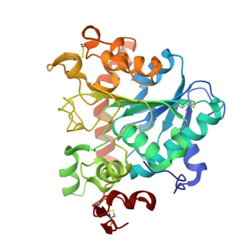
4ZV7 | pdb_00004zv7

Crystal structure of hexagonal form of lipase B from Candida antarctica


Experimental Data Snapshot

  • Method: X-RAY DIFFRACTION
  • Resolution: 2.00 Å
  • R-Value Free: 
    0.193 (Depositor), 0.204 (DCC) 
  • R-Value Work: 
    0.144 (Depositor), 0.157 (DCC) 
  • R-Value Observed: 
    0.147 (Depositor) 

Starting Model: experimental
View more details

wwPDB Validation 3D Report Full Report

Validation slider image for 4ZV7

This is version 2.2 of the entry. See complete history

Literature

Crystal and molecular structure of hexagonal form of lipase B from Candida antarctica.

Strzelczyk, P.Bujacz, G.D.Kiebasinski, P.Baszczyk, J.

(2016) Acta Biochim Pol 63: 103-109

  • DOI: https://doi.org/10.18388/abp.2015_1065
  • Primary Citation Related Structures: 
    4ZV7

  • PubMed Abstract: 

    During crystallization screenings of commercially available hydrolytic enzymes, the new, hexagonal crystal form of CAL-B, has been discovered and hereby reported. The NAG molecules, which were closing the glycosylation site in the orthorhombic form, in hexagonal structure make the glycosylation site open. It is unknown whether the opening and closing of the glycosylation site by the 'lid' NAG molecules, could be related to the opening and closing of the active center of the enzyme upon substrate binding and product release.


  • Organizational Affiliation
    • Lodz University of Technology, Institute of Technical Biochemistry, Łódź, Poland.

Macromolecule Content 

  • Total Structure Weight: 33.46 kDa 
  • Atom Count: 2,713 
  • Modeled Residue Count: 317 
  • Deposited Residue Count: 317 
  • Unique protein chains: 1

Macromolecules

Find similar proteins by:|  3D Structure
Entity ID: 1
MoleculeChains  Sequence LengthOrganismDetailsImage
Lipase B317Moesziomyces antarcticusMutation(s): 0 
EC: 3.1.1.3
UniProt
Find proteins for P41365 (Pseudozyma antarctica)
Explore P41365 
Go to UniProtKB:  P41365
Entity Groups
Sequence Clusters30% Identity50% Identity70% Identity90% Identity95% Identity100% Identity
UniProt GroupP41365
Glycosylation
Glycosylation Sites: 1
Sequence Annotations
Expand
Reference Sequence

Oligosaccharides

Help  
Entity ID: 2
MoleculeChains Length2D Diagram GlycosylationD Interactions
2-acetamido-2-deoxy-beta-D-glucopyranose-(1-4)-2-acetamido-2-deoxy-beta-D-glucopyranose
B
2N-Glycosylation
Glycosylation Resources
GlyTouCan: G42666HT
GlyCosmos: G42666HT
GlyGen: G42666HT

Experimental Data & Validation

Experimental Data

  • Method: X-RAY DIFFRACTION
  • Resolution: 2.00 Å
  • R-Value Free:  0.193 (Depositor), 0.204 (DCC) 
  • R-Value Work:  0.144 (Depositor), 0.157 (DCC) 
  • R-Value Observed: 0.147 (Depositor) 
Space Group: P 63 2 2
Unit Cell:
Length ( Å )Angle ( ˚ )
a = 89.034α = 90
b = 89.034β = 90
c = 137.257γ = 120
Software Package:
Software NamePurpose
REFMACrefinement
CrysalisProdata reduction
Aimlessdata scaling
PHASERphasing

Structure Validation

View Full Validation Report


Entry History 

Deposition Data

Revision History  (Full details and data files)

  • Version 1.0: 2016-01-13
    Type: Initial release
  • Version 1.1: 2016-03-30
    Changes: Database references
  • Version 1.2: 2017-09-13
    Changes: Data collection
  • Version 2.0: 2020-07-29
    Type: Remediation
    Reason: Carbohydrate remediation
    Changes: Advisory, Atomic model, Data collection, Derived calculations, Structure summary
  • Version 2.1: 2024-01-10
    Changes: Data collection, Database references, Refinement description, Structure summary
  • Version 2.2: 2024-11-13
    Changes: Structure summary