


Structures of a High-fidelity DNA Polymerase
Wang, W., Hellinga, H.W., Beese, L.S.To be published.
Experimental Data Snapshot
Starting Model: experimental
View more details
Entity ID: 1 | |||||
|---|---|---|---|---|---|
| Molecule | Chains | Sequence Length | Organism | Details | Image |
| DNA polymerase | A, B [auth D] | 592 | Geobacillus kaustophilus HTA426 | Mutation(s): 2 Gene Names: polA, GK2730 EC: 2.7.7.7 | 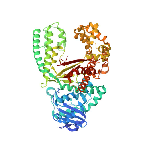 |
UniProt | |||||
Entity Groups | |||||
| Sequence Clusters | 30% Identity50% Identity70% Identity90% Identity95% Identity100% Identity | ||||
| UniProt Group | Q5KWC1 | ||||
Sequence AnnotationsExpand | |||||
Reference Sequence | |||||
Entity ID: 2 | ||||
| Molecule | Chains | Length | Organism | Image |
|---|---|---|---|---|
| DNA (5'-D(*CP*CP*TP*GP*AP*CP*TP*CP*(DDG))-3') | C [auth B], E | 9 | synthetic construct | 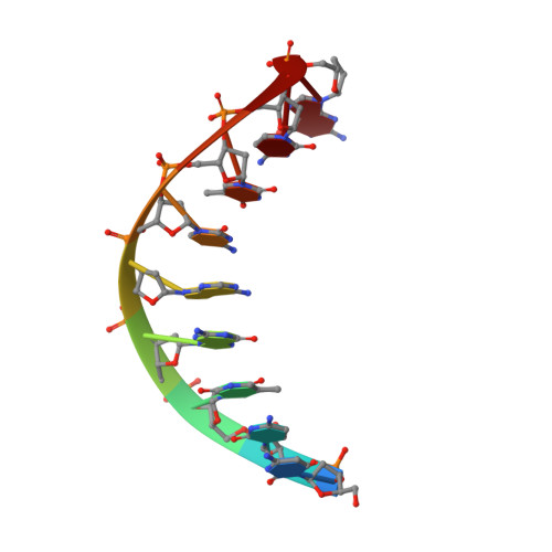 |
Sequence AnnotationsExpand | ||||
Reference Sequence | ||||
Entity ID: 3 | ||||
| Molecule | Chains | Length | Organism | Image |
|---|---|---|---|---|
| DNA (5'-D(*CP*AP*TP*TP*CP*GP*AP*GP*TP*CP*AP*GP*G)-3') | D [auth C], F | 13 | synthetic construct | 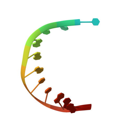 |
Sequence AnnotationsExpand | ||||
Reference Sequence | ||||
| Ligands 4 Unique | |||||
|---|---|---|---|---|---|
| ID | Chains | Name / Formula / InChI Key | 2D Diagram | 3D Interactions | |
| DG3 Download:Ideal Coordinates CCD File | H [auth D] | 2'-3'-DIDEOXYGUANOSINE-5'-TRIPHOSPHATE C10 H16 N5 O12 P3 HDRRAMINWIWTNU-NTSWFWBYSA-N |  | ||
| MPD Download:Ideal Coordinates CCD File | L [auth D] | (4S)-2-METHYL-2,4-PENTANEDIOL C6 H14 O2 SVTBMSDMJJWYQN-YFKPBYRVSA-N |  | ||
| SO4 Download:Ideal Coordinates CCD File | G [auth A], J [auth D], K [auth D] | SULFATE ION O4 S QAOWNCQODCNURD-UHFFFAOYSA-L |  | ||
| MN Download:Ideal Coordinates CCD File | I [auth D] | MANGANESE (II) ION Mn WAEMQWOKJMHJLA-UHFFFAOYSA-N |  | ||
| Length ( Å ) | Angle ( ˚ ) |
|---|---|
| a = 93.81 | α = 90 |
| b = 109.22 | β = 90 |
| c = 150.58 | γ = 90 |
| Software Name | Purpose |
|---|---|
| XSCALE | data scaling |
| PHENIX | refinement |
| PDB_EXTRACT | data extraction |
| XDS | data reduction |


