4YB5 | pdb_00004yb5

Adenosine triphosphate phosphoribosyltransferase from Campylobacter jejuni in complex with the allosteric inhibitor histidine


Experimental Data Snapshot

  • Method: X-RAY DIFFRACTION
  • Resolution: 2.24 Å
  • R-Value Free: 
    0.239 (Depositor), 0.227 (DCC) 
  • R-Value Work: 
    0.209 (Depositor) 
  • R-Value Observed: 
    0.210 (Depositor) 

Starting Model: experimental
View more details

wwPDB Validation 3D Report Full Report

Validation slider image for 4YB5

Ligand Structure Quality Assessment 


This is version 1.4 of the entry. See complete history

Literature

Campylobacter jejuni adenosine triphosphate phosphoribosyltransferase is an active hexamer that is allosterically controlled by the twisting of a regulatory tail.

Mittelstadt, G.Moggre, G.J.Panjikar, S.Nazmi, A.R.Parker, E.J.

(2016) Protein Sci 25: 1492-1506

  • DOI: https://doi.org/10.1002/pro.2948
  • Primary Citation Related Structures: 
    4YB5, 4YB6, 4YB7

  • PubMed Abstract: 

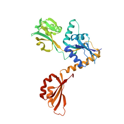
    Adenosine triphosphate phosphoribosyltransferase (ATP-PRT) catalyzes the first committed step of the histidine biosynthesis in plants and microorganisms. Here, we present the functional and structural characterization of the ATP-PRT from the pathogenic ε-proteobacteria Campylobacter jejuni (CjeATP-PRT). This enzyme is a member of the long form (HisGL ) ATP-PRT and is allosterically inhibited by histidine, which binds to a remote regulatory domain, and competitively inhibited by AMP. In the crystalline form, CjeATP-PRT was found to adopt two distinctly different hexameric conformations, with an open homohexameric structure observed in the presence of substrate ATP, and a more compact closed form present when inhibitor histidine is bound. CjeATP-PRT was observed to adopt only a hexameric quaternary structure in solution, contradicting previous hypotheses favoring an allosteric mechanism driven by an oligomer equilibrium. Instead, this study supports the conclusion that the ATP-PRT long form hexamer is the active species; the tightening of this structure in response to remote histidine binding results in an inhibited enzyme.


  • Organizational Affiliation
    • Maurice Wilkins Centre, Biomolecular Interaction Centre, Christchurch, 8140, New Zealand.

Macromolecule Content 

  • Total Structure Weight: 204.41 kDa 
  • Atom Count: 13,442 
  • Modeled Residue Count: 1,753 
  • Deposited Residue Count: 1,800 
  • Unique protein chains: 1

Macromolecules

Find similar proteins by:|  3D Structure
Entity ID: 1
MoleculeChains  Sequence LengthOrganismDetailsImage
ATP phosphoribosyltransferase
A, B, C, D, E
A, B, C, D, E, F
300Campylobacter jejuni RM1221Mutation(s): 0 
Gene Names: hisGCJE1769
EC: 2.4.2.17
UniProt
Find proteins for Q5HSJ4 (Campylobacter jejuni (strain RM1221))
Explore Q5HSJ4 
Go to UniProtKB:  Q5HSJ4
Entity Groups
Sequence Clusters30% Identity50% Identity70% Identity90% Identity95% Identity100% Identity
UniProt GroupQ5HSJ4
Sequence Annotations
Expand
Reference Sequence

Small Molecules

Ligands 5 Unique
IDChains Name / Formula / InChI Key2D Diagram3D Interactions
HIS

Query on HIS


Download:Ideal Coordinates CCD File
BA [auth F]
J [auth A]
N [auth B]
R [auth C]
U [auth D]
BA [auth F],
J [auth A],
N [auth B],
R [auth C],
U [auth D],
X [auth E]
HISTIDINE
C6 H10 N3 O2
HNDVDQJCIGZPNO-YFKPBYRVSA-O
PGE

Query on PGE


Download:Ideal Coordinates CCD File
P [auth C]TRIETHYLENE GLYCOL
C6 H14 O4
ZIBGPFATKBEMQZ-UHFFFAOYSA-N
PEG

Query on PEG


Download:Ideal Coordinates CCD File
AA [auth F],
T [auth D]
DI(HYDROXYETHYL)ETHER
C4 H10 O3
MTHSVFCYNBDYFN-UHFFFAOYSA-N
SCN

Query on SCN


Download:Ideal Coordinates CCD File
G [auth A]
H [auth A]
L [auth B]
O [auth C]
V [auth E]
G [auth A],
H [auth A],
L [auth B],
O [auth C],
V [auth E],
W [auth E],
Y [auth F],
Z [auth F]
THIOCYANATE ION
C N S
ZMZDMBWJUHKJPS-UHFFFAOYSA-M
K

Query on K


Download:Ideal Coordinates CCD File
I [auth A],
K [auth A],
M [auth B],
Q [auth C],
S [auth C]
POTASSIUM ION
K
NPYPAHLBTDXSSS-UHFFFAOYSA-N

Experimental Data & Validation

Experimental Data

  • Method: X-RAY DIFFRACTION
  • Resolution: 2.24 Å
  • R-Value Free:  0.239 (Depositor), 0.227 (DCC) 
  • R-Value Work:  0.209 (Depositor) 
  • R-Value Observed: 0.210 (Depositor) 
Space Group: P 1 21 1
Unit Cell:
Length ( Å )Angle ( ˚ )
a = 91.137α = 90
b = 123.216β = 110.66
c = 95.701γ = 90
Software Package:
Software NamePurpose
REFMACrefinement
XDSdata reduction
Aimlessdata scaling
PHASERphasing
MOLREPphasing

Structure Validation

View Full Validation Report


Ligand Structure Quality Assessment 


Entry History 

Deposition Data

Revision History  (Full details and data files)

  • Version 1.0: 2016-03-09
    Type: Initial release
  • Version 1.1: 2017-09-27
    Changes: Author supporting evidence, Data collection, Derived calculations
  • Version 1.2: 2017-11-01
    Changes: Author supporting evidence
  • Version 1.3: 2018-09-05
    Changes: Data collection, Database references
  • Version 1.4: 2023-09-27
    Changes: Data collection, Database references, Derived calculations, Refinement description