5A0R

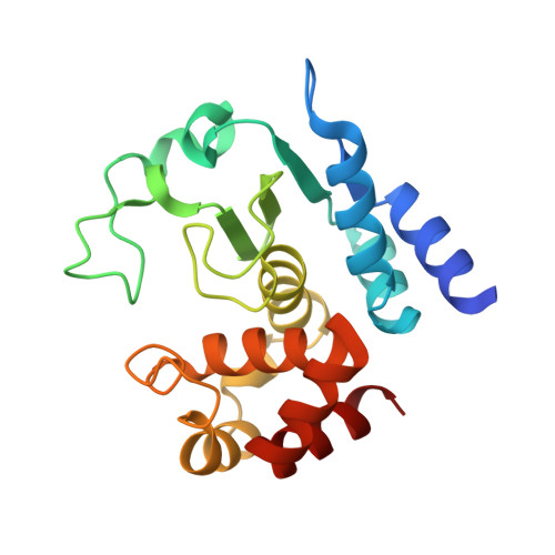
Product peptide-bound structure of metalloprotease Zmp1 variant E143A from Clostridium difficile


Experimental Data Snapshot

  • Method: X-RAY DIFFRACTION
  • Resolution: 1.25 Å
  • R-Value Free: 0.162 
  • R-Value Work: 0.141 
  • R-Value Observed: 0.142 

Starting Model: experimental
View more details

wwPDB Validation   3D Report Full Report


This is version 2.2 of the entry. See complete history


Literature

Structural Basis of Proline-Proline Peptide Bond Specificity of the Metalloprotease Zmp1 Implicated in Motility of Clostridium Difficile.

Schacherl, M.Pichlo, C.Neundorf, I.Baumann, U.

(2015) Structure 23: 1632

  • DOI: https://doi.org/10.1016/j.str.2015.06.018
  • Primary Citation of Related Structures:  
    5A0P, 5A0R, 5A0S, 5A0X

  • PubMed Abstract: 

    Clostridium difficile is a pathogenic bacterium causing gastrointestinal diseases from mild diarrhea to toxic megacolon. In common with other pathogenic bacteria, C. difficile secretes proteins involved in adhesion, colonization, and dissemination. The recently identified Zmp1 is an extracellular metalloprotease showing a unique specificity for Pro-Pro peptide bonds. The endogenous substrates of Zmp1 are two surface proteins implicated in adhesion of C. difficile to surface proteins of human cells. Thus, Zmp1 is believed to be involved in the regulation of the adhesion-motility balance of C. difficile. Here, we report crystal structures of Zmp1 from C. difficile in its unbound and peptide-bound forms. The structure analysis revealed a fold similar to Bacillus anthracis lethal factor. Crystal structures in the open and closed conformation of the S-loop shed light on the mode of binding of the substrate, and reveal important residues for substrate recognition and the strict specificity of Zmp1 for Pro-Pro peptide bonds.


  • Organizational Affiliation

    Institute of Biochemistry, University of Cologne, 50674 Cologne, Germany. Electronic address: magdalena.schacherl@uni-koeln.de.


Macromolecules
Find similar proteins by:  (by identity cutoff)  |  3D Structure
Entity ID: 1
MoleculeChains Sequence LengthOrganismDetailsImage
ZINC METALLOPROTEASE ZMP1
A, B
198Clostridioides difficile 630Mutation(s): 1 
EC: 3.4.24 (PDB Primary Data), 3.4.24.89 (UniProt)
UniProt
Find proteins for Q183R7 (Clostridioides difficile (strain 630))
Explore Q183R7 
Go to UniProtKB:  Q183R7
Entity Groups  
Sequence Clusters30% Identity50% Identity70% Identity90% Identity95% Identity100% Identity
UniProt GroupQ183R7
Sequence Annotations
Expand
  • Reference Sequence

Find similar proteins by:  Sequence   |   3D Structure  

Entity ID: 2
MoleculeChains Sequence LengthOrganismDetailsImage
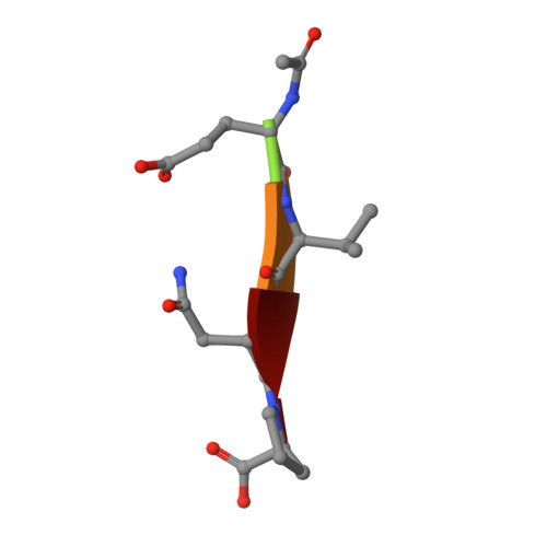
PRODUCT PEPTIDEC [auth D],
D [auth E]
5synthetic constructMutation(s): 0 
Sequence Annotations
Expand
  • Reference Sequence
Experimental Data & Validation

Experimental Data

  • Method: X-RAY DIFFRACTION
  • Resolution: 1.25 Å
  • R-Value Free: 0.162 
  • R-Value Work: 0.141 
  • R-Value Observed: 0.142 
  • Space Group: P 1 21 1
Unit Cell:
Length ( Å )Angle ( ˚ )
a = 37.06α = 90
b = 42.96β = 92.54
c = 119.21γ = 90
Software Package:
Software NamePurpose
PHENIXrefinement
XDSdata reduction
XSCALEdata scaling
PHASERphasing

Structure Validation

View Full Validation Report


Entry History 

Deposition Data

Revision History  (Full details and data files)

  • Version 1.0: 2015-08-05
    Type: Initial release
  • Version 1.1: 2015-09-16
    Changes: Database references
  • Version 1.2: 2019-03-06
    Changes: Data collection, Derived calculations, Experimental preparation, Other
  • Version 2.0: 2019-10-23
    Changes: Atomic model, Data collection, Other
  • Version 2.1: 2024-01-10
    Changes: Data collection, Database references, Derived calculations, Refinement description
  • Version 2.2: 2024-11-13
    Changes: Structure summary