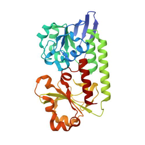
5A1J | pdb_00005a1j

Periplasmic Binding Protein CeuE in complex with ferric 4-LICAM


Experimental Data Snapshot

  • Method: X-RAY DIFFRACTION
  • Resolution: 1.60 Å
  • R-Value Free: 
    0.312 (Depositor), 0.310 (DCC) 
  • R-Value Work: 
    0.264 (Depositor), 0.270 (DCC) 
  • R-Value Observed: 
    0.267 (Depositor) 

Starting Model: experimental
View more details

wwPDB Validation   3D Report Full Report


Ligand Structure Quality Assessment 

Created with Raphaël 2.3.0Worse 01 BetterLigand structure goodness of fit to experimental dataBest fitted LCMClick on this verticalbar to view details

This is version 1.1 of the entry. See complete history


Literature

Interactions of a Periplasmic Binding Protein with a Tetradentate Siderophore Mimic.

Raines, D.J.Moroz, O.V.Wilson, K.S.Duhme-Klair, A.

(2013) Angew Chem Int Ed Engl 52: 4595

  • DOI: https://doi.org/10.1002/anie.201300751
  • Primary Citation of Related Structures:  
    3ZKW, 5A1J

  • PubMed Abstract: 

    Iron-bound structure: The ferric complex of a tetradentate siderophore mimic was synthesized and co-crystallized with the periplasmic binding protein CeuE of Campylobacter jejuni. In addition to electrostatic and hydrogen-bonding interactions between the binding pocket and the substrate, the structure showed direct coordination of two amino acid side chains to the Fe(III) center (orange, see figure).


  • Organizational Affiliation

    Department of Chemistry, University of York, Heslington, York, YO10 5DD, UK.


Macromolecules
Find similar proteins by:  (by identity cutoff)  |  3D Structure
Entity ID: 1
MoleculeChains Sequence LengthOrganismDetailsImage
ENTEROCHELIN UPTAKE PERIPLASMIC BINDING PROTEIN288Campylobacter jejuniMutation(s): 0 
UniProt
Find proteins for Q0P8Q4 (Campylobacter jejuni subsp. jejuni serotype O:2 (strain ATCC 700819 / NCTC 11168))
Explore Q0P8Q4 
Go to UniProtKB:  Q0P8Q4
Entity Groups  
Sequence Clusters30% Identity50% Identity70% Identity90% Identity95% Identity100% Identity
UniProt GroupQ0P8Q4
Sequence Annotations
Expand
  • Reference Sequence
Small Molecules
Experimental Data & Validation

Experimental Data

  • Method: X-RAY DIFFRACTION
  • Resolution: 1.60 Å
  • R-Value Free:  0.312 (Depositor), 0.310 (DCC) 
  • R-Value Work:  0.264 (Depositor), 0.270 (DCC) 
  • R-Value Observed: 0.267 (Depositor) 
Space Group: P 21 21 21
Unit Cell:
Length ( Å )Angle ( ˚ )
a = 60.42α = 90
b = 66.89β = 90
c = 67.3γ = 90
Software Package:
Software NamePurpose
REFMACrefinement
xia2data reduction
MOLREPphasing

Structure Validation

View Full Validation Report


Ligand Structure Quality Assessment 

Created with Raphaël 2.3.0Worse 01 BetterLigand structure goodness of fit to experimental dataBest fitted LCMClick on this verticalbar to view details

Entry History 

Deposition Data

Revision History  (Full details and data files)

  • Version 1.0: 2015-05-13
    Type: Initial release
  • Version 1.1: 2024-01-10
    Changes: Data collection, Database references, Derived calculations, Other, Refinement description