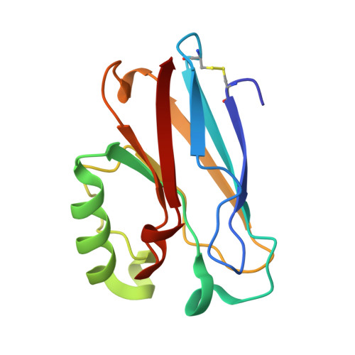
5AZU

CRYSTAL STRUCTURE ANALYSIS OF OXIDIZED PSEUDOMONAS AERUGINOSA AZURIN AT PH 5.5 AND PH 9.0. A PH-INDUCED CONFORMATIONAL TRANSITION INVOLVES A PEPTIDE BOND FLIP


Experimental Data Snapshot

  • Method: X-RAY DIFFRACTION
  • Resolution: 1.90 Å

wwPDB Validation   3D Report Full Report


This is version 1.4 of the entry. See complete history


Literature

Crystal structure analysis of oxidized Pseudomonas aeruginosa azurin at pH 5.5 and pH 9.0. A pH-induced conformational transition involves a peptide bond flip.

Nar, H.Messerschmidt, A.Huber, R.van de Kamp, M.Canters, G.W.

(1991) J Mol Biol 221: 765-772

  • DOI: https://doi.org/10.1016/0022-2836(91)80173-r
  • Primary Citation of Related Structures:  
    4AZU, 5AZU

  • PubMed Abstract: 

    The X-ray crystal structure of recombinant wild-type azurin from Pseudomonas aeruginosa was determined by difference Fourier techniques using phases derived from the structure of the mutant His35Leu. Two data sets were collected from a single crystal of oxidized azurin soaked in mother liquor buffered at pH 5.5 and pH 9.0, respectively. Both data sets extend to 1.93 A resolution. The two pH forms were refined independently to crystallographic R-factors of 17.6% (pH 5.5) and 17.5% (pH 9.0). The conformational transition previously attributed to the protonation/deprotonation of residue His35 (pKa(red) = 7.3, pKa(ox) = 6.2), which lies in a crevice of the protein close to the copper binding site, involves a concomitant Pro36-Gly37 main-chain peptide bond flip. At the lower pH, the protonated imidazole N delta 1 of His35 forms a strong hydrogen bond with the carbonyl oxygen from Pro36, while at alkaline pH the deprotonated N delta 1 acts as an acceptor of a weak hydrogen bond from HN Gly37. The structure of the remainder of the azurin molecule, including the copper binding site, is not significantly affected by this transition.


  • Organizational Affiliation

    Max Planck Institut für Biochemie, Martinsried bei München, Germany.


Macromolecules
Find similar proteins by:  (by identity cutoff)  |  3D Structure
Entity ID: 1
MoleculeChains Sequence LengthOrganismDetailsImage
AZURIN
A, B, C, D
128Pseudomonas aeruginosaMutation(s): 0 
UniProt
Find proteins for P00282 (Pseudomonas aeruginosa (strain ATCC 15692 / DSM 22644 / CIP 104116 / JCM 14847 / LMG 12228 / 1C / PRS 101 / PAO1))
Explore P00282 
Go to UniProtKB:  P00282
Entity Groups  
Sequence Clusters30% Identity50% Identity70% Identity90% Identity95% Identity100% Identity
UniProt GroupP00282
Sequence Annotations
Expand
  • Reference Sequence
Experimental Data & Validation

Experimental Data

  • Method: X-RAY DIFFRACTION
  • Resolution: 1.90 Å
  • Space Group: P 21 21 21
Unit Cell:
Length ( Å )Angle ( ˚ )
a = 57.76α = 90
b = 81.09β = 90
c = 109.94γ = 90
Software Package:
Software NamePurpose
X-PLORmodel building
X-PLORrefinement
X-PLORphasing

Structure Validation

View Full Validation Report


Entry History 

Deposition Data

Revision History  (Full details and data files)

  • Version 1.0: 1994-01-31
    Type: Initial release
  • Version 1.1: 2008-03-25
    Changes: Version format compliance
  • Version 1.2: 2011-07-13
    Changes: Version format compliance
  • Version 1.3: 2024-06-05
    Changes: Data collection, Database references, Derived calculations, Other
  • Version 1.4: 2024-10-30
    Changes: Structure summary