


Crystal structure and ligand affinity of avidin in the complex with 4-hydroxyazobenzene-2-carboxylic acid
Strzelczyk, P., Bujacz, G.(2016) J Mol Struct 1109: 232-238
Experimental Data Snapshot
Starting Model: experimental
View more details
(2016) J Mol Struct 1109: 232-238
Entity ID: 1 | |||||
|---|---|---|---|---|---|
| Molecule | Chains | Sequence Length | Organism | Details | Image |
| Avidin | 128 | Gallus gallus | Mutation(s): 0 | 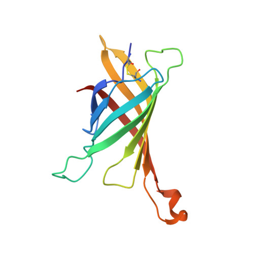 | |
UniProt | |||||
Entity Groups | |||||
| Sequence Clusters | 30% Identity50% Identity70% Identity90% Identity95% Identity100% Identity | ||||
| UniProt Group | P02701 | ||||
Glycosylation | |||||
| Glycosylation Sites: 1 | |||||
Sequence AnnotationsExpand | |||||
Reference Sequence | |||||
| Ligands 2 Unique | |||||
|---|---|---|---|---|---|
| ID | Chains | Name / Formula / InChI Key | 2D Diagram | 3D Interactions | |
| 55R Download:Ideal Coordinates CCD File | C [auth A] | 2-[2-(4-oxocyclohexa-2,5-dien-1-ylidene)hydrazinyl]benzoic acid C13 H10 N2 O3 FBVSMDPNVYJNON-UHFFFAOYSA-N |  | ||
| NAG Download:Ideal Coordinates CCD File | B [auth A] | 2-acetamido-2-deoxy-beta-D-glucopyranose C8 H15 N O6 OVRNDRQMDRJTHS-FMDGEEDCSA-N |  | ||
| Length ( Å ) | Angle ( ˚ ) |
|---|---|
| a = 61.31 | α = 90 |
| b = 61.31 | β = 90 |
| c = 120.53 | γ = 120 |
| Software Name | Purpose |
|---|---|
| REFMAC | refinement |
| XDS | data reduction |
| XDS | data scaling |
| MOLREP | phasing |
| Funding Organization | Location | Grant Number |
|---|---|---|
| National Science Centre | Poland | DEC-2013/11/N/ST5/01296 |


