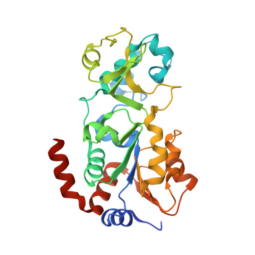
5D7N

Crystal structure of human Sirt3 at an improved resolution


Experimental Data Snapshot

  • Method: X-RAY DIFFRACTION
  • Resolution: 1.83 Å
  • R-Value Free: 0.203 
  • R-Value Work: 0.175 
  • R-Value Observed: 0.176 

Starting Model: experimental
View more details

wwPDB Validation   3D Report Full Report


This is version 1.2 of the entry. See complete history


Literature

Seeding for sirtuins: microseed matrix seeding to obtain crystals of human Sirt3 and Sirt2 suitable for soaking.

Rumpf, T.Gerhardt, S.Einsle, O.Jung, M.

(2015) Acta Crystallogr F Struct Biol Commun 71: 1498-1510

  • DOI: https://doi.org/10.1107/S2053230X15019986
  • Primary Citation of Related Structures:  
    5D7N, 5D7O, 5D7P, 5D7Q

  • PubMed Abstract: 

    Sirtuins constitute a family of NAD(+)-dependent enzymes that catalyse the cleavage of various acyl groups from the ℇ-amino group of lysines. They regulate a series of cellular processes and their misregulation has been implicated in various diseases, making sirtuins attractive drug targets. To date, only a few sirtuin modulators have been reported that are suitable for cellular research and their development has been hampered by a lack of structural information. In this work, microseed matrix seeding (MMS) was used to obtain crystals of human Sirt3 in its apo form and of human Sirt2 in complex with ADP ribose (ADPR). Crystal formation using MMS was predictable, less error-prone and yielded a higher number of crystals per drop than using conventional crystallization screening methods. The crystals were used to solve the crystal structures of apo Sirt3 and of Sirt2 in complex with ADPR at an improved resolution, as well as the crystal structures of Sirt2 in complex with ADPR and the indoles EX527 and CHIC35. These Sirt2-ADPR-indole complexes unexpectedly contain two indole molecules and provide novel insights into selective Sirt2 inhibition. The MMS approach for Sirt2 and Sirt3 may be used as the basis for structure-based optimization of Sirt2/3 inhibitors in the future.


  • Organizational Affiliation

    Institute of Pharmaceutical Sciences, Albert-Ludwigs-University Freiburg, Albertstrasse 25, 79104 Freiburg, Baden-Württemberg, Germany.


Macromolecules
Find similar proteins by:  (by identity cutoff)  |  3D Structure
Entity ID: 1
MoleculeChains Sequence LengthOrganismDetailsImage
NAD-dependent protein deacetylase sirtuin-3, mitochondrial
A, B, C, D, E
A, B, C, D, E, F
281Homo sapiensMutation(s): 0 
Gene Names: SIRT3SIR2L3
EC: 3.5.1 (PDB Primary Data), 2.3.1.286 (UniProt)
UniProt & NIH Common Fund Data Resources
Find proteins for Q9NTG7 (Homo sapiens)
Explore Q9NTG7 
Go to UniProtKB:  Q9NTG7
PHAROS:  Q9NTG7
GTEx:  ENSG00000142082 
Entity Groups  
Sequence Clusters30% Identity50% Identity70% Identity90% Identity95% Identity100% Identity
UniProt GroupQ9NTG7
Sequence Annotations
Expand
  • Reference Sequence
Small Molecules
Ligands 7 Unique
IDChains Name / Formula / InChI Key2D Diagram3D Interactions
1PE
Query on 1PE

Download Ideal Coordinates CCD File 
R [auth D]PENTAETHYLENE GLYCOL
C10 H22 O6
JLFNLZLINWHATN-UHFFFAOYSA-N
PG4
Query on PG4

Download Ideal Coordinates CCD File 
W [auth F]TETRAETHYLENE GLYCOL
C8 H18 O5
UWHCKJMYHZGTIT-UHFFFAOYSA-N
PGE
Query on PGE

Download Ideal Coordinates CCD File 
U [auth E]TRIETHYLENE GLYCOL
C6 H14 O4
ZIBGPFATKBEMQZ-UHFFFAOYSA-N
PEG
Query on PEG

Download Ideal Coordinates CCD File 
H [auth A]
I [auth A]
J [auth A]
M [auth B]
O [auth C]
H [auth A],
I [auth A],
J [auth A],
M [auth B],
O [auth C],
Q [auth D]
DI(HYDROXYETHYL)ETHER
C4 H10 O3
MTHSVFCYNBDYFN-UHFFFAOYSA-N
GOL
Query on GOL

Download Ideal Coordinates CCD File 
S [auth D]GLYCEROL
C3 H8 O3
PEDCQBHIVMGVHV-UHFFFAOYSA-N
ZN
Query on ZN

Download Ideal Coordinates CCD File 
G [auth A]
L [auth B]
N [auth C]
P [auth D]
T [auth E]
G [auth A],
L [auth B],
N [auth C],
P [auth D],
T [auth E],
V [auth F]
ZINC ION
Zn
PTFCDOFLOPIGGS-UHFFFAOYSA-N
MG
Query on MG

Download Ideal Coordinates CCD File 
K [auth A]MAGNESIUM ION
Mg
JLVVSXFLKOJNIY-UHFFFAOYSA-N
Experimental Data & Validation

Experimental Data

  • Method: X-RAY DIFFRACTION
  • Resolution: 1.83 Å
  • R-Value Free: 0.203 
  • R-Value Work: 0.175 
  • R-Value Observed: 0.176 
  • Space Group: P 1 21 1
Unit Cell:
Length ( Å )Angle ( ˚ )
a = 84.99α = 90
b = 143.85β = 116.33
c = 89.46γ = 90
Software Package:
Software NamePurpose
REFMACrefinement
MOLREPphasing
Aimlessdata scaling
XDSdata reduction

Structure Validation

View Full Validation Report


Entry History & Funding Information

Deposition Data


Funding OrganizationLocationGrant Number
German Research FoundationGermanyJu295/8-1
German Research FoundationGermanySFB992 Medical Epigenetics, Project Z02

Revision History  (Full details and data files)

  • Version 1.0: 2015-12-02
    Type: Initial release
  • Version 1.1: 2015-12-09
    Changes: Database references
  • Version 1.2: 2024-01-10
    Changes: Author supporting evidence, Data collection, Database references, Derived calculations, Refinement description