Crystal structure of unliganded Escherichia coli dihydrofolate reductase. Ligand-induced conformational changes and cooperativity in binding.
Bystroff, C., Kraut, J.(1991) Biochemistry 30: 2227-2239
- PubMed: 1998681 Search on PubMed
- DOI: https://doi.org/10.1021/bi00222a028
- Primary Citation Related Structures:
1DRA, 1DRB, 5DFR - PubMed Abstract:
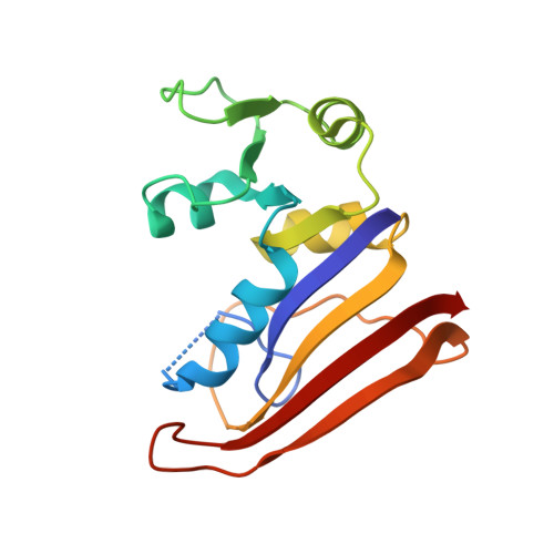
The crystal structure of unliganded dihydrofolate reductase (DHFR) from Escherichia coli has been solved and refined to an R factor of 19% at 2.3-A resolution in a crystal form that is nonisomorphous with each of the previously reported E. coli DHFR crystal structures [Bolin, J. T., Filman, D. J., Matthews, D. A., Hamlin, B. C., & Kraut, J. (1982) J. Biol. Chem. 257, 13650-13662; Bystroff, C., Oatley, S. J., & Kraut, J. (1990) Biochemistry 29, 3263-3277]. Significant conformational changes occur between the apoenzyme and each of the complexes: the NADP+ holoenzyme, the folate-NADP+ ternary complex, and the methotrexate (MTX) binary complex. The changes are small, with the largest about 3 A and most of them less than 1 A. For simplicity a two-domain description is adopted in which one domain contains the NADP+ 2'-phosphate binding site and the binding sites for the rest of the coenzyme and for the substrate lie between the two domains. Binding of either NADP+ or MTX induces a closing of the PABG-binding cleft and realignment of alpha-helices C and F which bind the pyrophosphate of the coenzyme. Formation of the ternary complex from the holoenzyme does not involve further relative domain shifts but does involve a shift of alpha-helix B and a floppy loop (the Met-20 loop) that precedes alpha B. These observations suggest a mechanism for cooperativity in binding between substrate and coenzyme wherein the greatest degree of cooperativity is expressed in the transition-state complex. We explore the idea that the MTX binary complex in some ways resembles the transition-state complex.
- Department of Chemistry, University of California, San Diego, La Jolla 92093.
Organizational Affiliation: