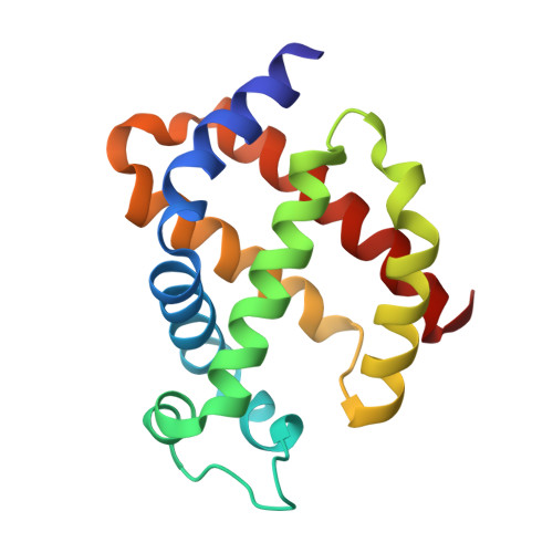
5EOH

Crystal structure of murine neuroglobin at 270 MPa pressure


Experimental Data Snapshot

  • Method: X-RAY DIFFRACTION
  • Resolution: 1.90 Å
  • R-Value Free: 0.221 
  • R-Value Work: 0.169 
  • R-Value Observed: 0.171 

Starting Model: experimental
View more details

wwPDB Validation   3D Report Full Report


Ligand Structure Quality Assessment 


This is version 1.3 of the entry. See complete history


Literature

Determinants of neuroglobin plasticity highlighted by joint coarse-grained simulations and high pressure crystallography.

Colloc'h, N.Sacquin-Mora, S.Avella, G.Dhaussy, A.C.Prange, T.Vallone, B.Girard, E.

(2017) Sci Rep 7: 1858-1858

  • DOI: https://doi.org/10.1038/s41598-017-02097-1
  • Primary Citation of Related Structures:  
    5EET, 5EOH, 5EQM, 5EU2, 5EV5, 5EYJ, 5EYS, 5F0B, 5F2A

  • PubMed Abstract: 

    Investigating the effect of pressure sheds light on the dynamics and plasticity of proteins, intrinsically correlated to functional efficiency. Here we detail the structural response to pressure of neuroglobin (Ngb), a hexacoordinate globin likely to be involved in neuroprotection. In murine Ngb, reversible coordination is achieved by repositioning the heme more deeply into a large internal cavity, the "heme sliding mechanism". Combining high pressure crystallography and coarse-grain simulations on wild type Ngb as well as two mutants, one (V101F) with unaffected and another (F106W) with decreased affinity for CO, we show that Ngb hinges around a rigid mechanical nucleus of five hydrophobic residues (V68, I72, V109, L113, Y137) during its conformational transition induced by gaseous ligand, that the intrinsic flexibility of the F-G loop appears essential to drive the heme sliding mechanism, and that residue Val 101 may act as a sensor of the interaction disruption between the heme and the distal histidine.


  • Organizational Affiliation

    ISTCT CNRS UNICAEN CEA Normandie Univ., CERVOxy team, centre Cyceron, 14000, Caen, France. colloch@cyceron.fr.


Macromolecules
Find similar proteins by:  (by identity cutoff)  |  3D Structure
Entity ID: 1
MoleculeChains Sequence LengthOrganismDetailsImage
Neuroglobin148Mus musculusMutation(s): 2 
Gene Names: Ngb
EC: 1.7
UniProt
Find proteins for Q9ER97 (Mus musculus)
Explore Q9ER97 
Go to UniProtKB:  Q9ER97
Entity Groups  
Sequence Clusters30% Identity50% Identity70% Identity90% Identity95% Identity100% Identity
UniProt GroupQ9ER97
Sequence Annotations
Expand
  • Reference Sequence
Experimental Data & Validation

Experimental Data

  • Method: X-RAY DIFFRACTION
  • Resolution: 1.90 Å
  • R-Value Free: 0.221 
  • R-Value Work: 0.169 
  • R-Value Observed: 0.171 
  • Space Group: H 3 2
Unit Cell:
Length ( Å )Angle ( ˚ )
a = 87.56α = 90
b = 87.56β = 90
c = 113.636γ = 120
Software Package:
Software NamePurpose
REFMACrefinement
SCALAdata scaling
PHASERphasing
DMphasing
PDB_EXTRACTdata extraction

Structure Validation

View Full Validation Report


Ligand Structure Quality Assessment 


Entry History 

Deposition Data

Revision History  (Full details and data files)

  • Version 1.0: 2016-10-19
    Type: Initial release
  • Version 1.1: 2017-05-31
    Changes: Database references
  • Version 1.2: 2021-07-28
    Changes: Structure summary
  • Version 1.3: 2024-01-10
    Changes: Data collection, Database references, Refinement description