5F1U | pdb_00005f1u

biomimetic design results in a potent allosteric inhibitor of dihydrodipicolinate synthase from Campylobacter jejuni


Experimental Data Snapshot

  • Method: X-RAY DIFFRACTION
  • Resolution: 2.35 Å
  • R-Value Free: 
    0.229 (Depositor), 0.228 (DCC) 
  • R-Value Work: 
    0.171 (Depositor), 0.177 (DCC) 
  • R-Value Observed: 
    0.173 (Depositor) 

Starting Model: experimental
View more details

wwPDB Validation 3D Report Full Report

Validation slider image for 5F1U

Ligand Structure Quality Assessment 


This is version 1.3 of the entry. See complete history

Literature

Biomimetic Design Results in a Potent Allosteric Inhibitor of Dihydrodipicolinate Synthase from Campylobacter jejuni.

Skovpen, Y.V.Conly, C.J.Sanders, D.A.Palmer, D.R.

(2016) J Am Chem Soc 138: 2014-2020

  • DOI: https://doi.org/10.1021/jacs.5b12695
  • Primary Citation Related Structures: 
    5F1U, 5F1V

  • PubMed Abstract: 

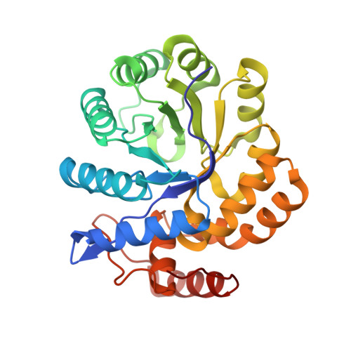
    Dihydrodipicolinate synthase (DHDPS), an enzyme required for bacterial peptidoglycan biosynthesis, catalyzes the condensation of pyruvate and β-aspartate semialdehyde (ASA) to form a cyclic product which dehydrates to form dihydrodipicolinate. DHDPS has, for several years, been considered a putative target for novel antibiotics. We have designed the first potent inhibitor of this enzyme by mimicking its natural allosteric regulation by lysine, and obtained a crystal structure of the protein-inhibitor complex at 2.2 Å resolution. This novel inhibitor, which we named "bislysine", resembles two lysine molecules linked by an ethylene bridge between the α-carbon atoms. Bislysine is a mixed partial inhibitor with respect to the first substrate, pyruvate, and a noncompetitive partial inhibitor with respect to ASA, and binds to all forms of the enzyme with a Ki near 200 nM, more than 300 times more tightly than lysine. Hill plots show that the inhibition is cooperative, indicating that the allosteric sites are not independent despite being located on opposite sides of the protein tetramer, separated by approximately 50 Å. A mutant enzyme resistant to lysine inhibition, Y110F, is strongly inhibited by this novel inhibitor, suggesting this may be a promising strategy for antibiotic development.


  • Organizational Affiliation
    • Department of Chemistry, University of Saskatchewan , 110 Science Place, Saskatoon, Saskatchewan, Canada S7N 5C9.

Macromolecule Content 

  • Total Structure Weight: 131.89 kDa 
  • Atom Count: 9,507 
  • Modeled Residue Count: 1,184 
  • Deposited Residue Count: 1,188 
  • Unique protein chains: 1

Macromolecules

Find similar proteins by:|  3D Structure
Entity ID: 1
MoleculeChains  Sequence LengthOrganismDetailsImage
4-hydroxy-tetrahydrodipicolinate synthase
A, B, C, D
297Campylobacter jejuniMutation(s): 1 
Gene Names: dapA
EC: 4.3.3.7
UniProt
Find proteins for Q9PPB4 (Campylobacter jejuni subsp. jejuni serotype O:2 (strain ATCC 700819 / NCTC 11168))
Explore Q9PPB4 
Go to UniProtKB:  Q9PPB4
Entity Groups
Sequence Clusters30% Identity50% Identity70% Identity90% Identity95% Identity100% Identity
UniProt GroupQ9PPB4
Sequence Annotations
Expand
Reference Sequence

Small Molecules

Ligands 5 Unique
IDChains Name / Formula / InChI Key2D Diagram3D Interactions
3VN

Query on 3VN


Download:Ideal Coordinates CCD File
E [auth A],
Q [auth D]
(2R,5R)-2,5-diamino-2,5-bis(4-aminobutyl)hexanedioic acid
C14 H30 N4 O4
XYUHPPVEDOOPDE-ZIAGYGMSSA-N
PG4

Query on PG4


Download:Ideal Coordinates CCD File
F [auth A]TETRAETHYLENE GLYCOL
C8 H18 O5
UWHCKJMYHZGTIT-UHFFFAOYSA-N
PGE

Query on PGE


Download:Ideal Coordinates CCD File
K [auth B]TRIETHYLENE GLYCOL
C6 H14 O4
ZIBGPFATKBEMQZ-UHFFFAOYSA-N
EDO

Query on EDO


Download:Ideal Coordinates CCD File
G [auth A]
H [auth A]
I [auth A]
L [auth B]
M [auth B]
G [auth A],
H [auth A],
I [auth A],
L [auth B],
M [auth B],
N [auth B],
O [auth C],
P [auth C],
R [auth D],
S [auth D]
1,2-ETHANEDIOL
C2 H6 O2
LYCAIKOWRPUZTN-UHFFFAOYSA-N
CL

Query on CL


Download:Ideal Coordinates CCD File
J [auth B]CHLORIDE ION
Cl
VEXZGXHMUGYJMC-UHFFFAOYSA-M

Experimental Data & Validation

Experimental Data

  • Method: X-RAY DIFFRACTION
  • Resolution: 2.35 Å
  • R-Value Free:  0.229 (Depositor), 0.228 (DCC) 
  • R-Value Work:  0.171 (Depositor), 0.177 (DCC) 
  • R-Value Observed: 0.173 (Depositor) 
Space Group: P 21 21 21
Unit Cell:
Length ( Å )Angle ( ˚ )
a = 79.59α = 90
b = 96.78β = 90
c = 145.98γ = 90
Software Package:
Software NamePurpose
XSCALEdata scaling
PDB_EXTRACTdata extraction
PHENIXrefinement
XDSdata reduction
MOLREPphasing

Structure Validation

View Full Validation Report


Ligand Structure Quality Assessment 


Entry History 

Deposition Data

Revision History  (Full details and data files)

  • Version 1.0: 2016-02-17
    Type: Initial release
  • Version 1.1: 2016-02-24
    Changes: Database references
  • Version 1.2: 2017-09-27
    Changes: Author supporting evidence, Derived calculations
  • Version 1.3: 2023-09-27
    Changes: Data collection, Database references, Refinement description