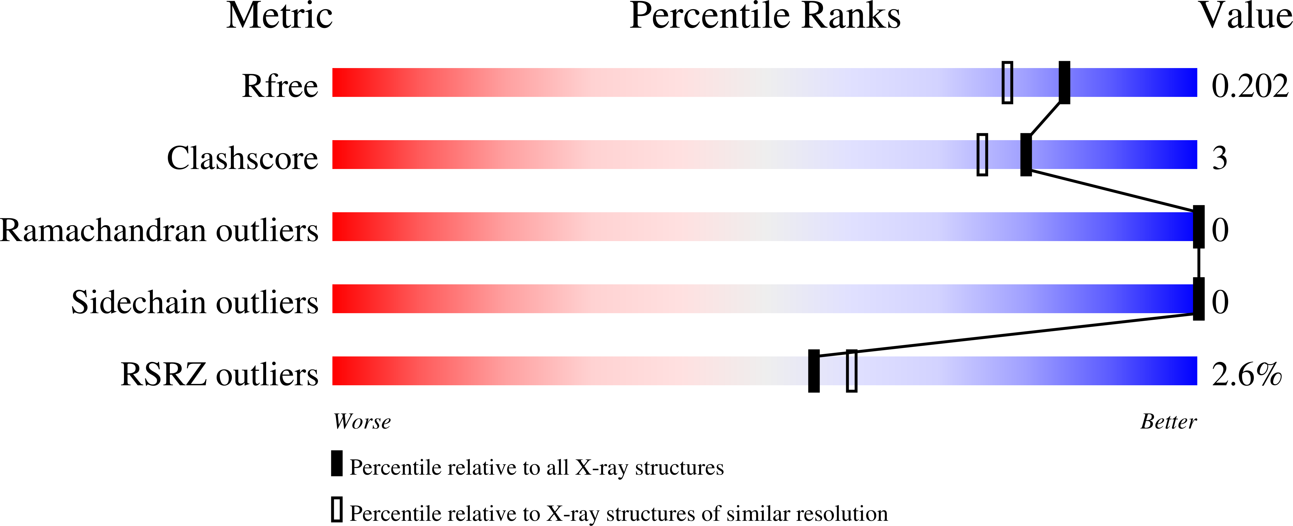
5F6W

Crystal structure of Ubc9 (K48/K49A/E54A) complexed with Fragment 1 (biphenol)


Experimental Data Snapshot

  • Method: X-RAY DIFFRACTION
  • Resolution: 1.70 Å
  • R-Value Free: 0.200 
  • R-Value Work: 0.172 
  • R-Value Observed: 0.174 

Starting Model: experimental
View more details

wwPDB Validation   3D Report Full Report


Ligand Structure Quality Assessment 


This is version 1.2 of the entry. See complete history


Literature

Insights Into the Allosteric Inhibition of the SUMO E2 Enzyme Ubc9.

Hewitt, W.M.Lountos, G.T.Zlotkowski, K.Dahlhauser, S.D.Saunders, L.B.Needle, D.Tropea, J.E.Zhan, C.Wei, G.Ma, B.Nussinov, R.Waugh, D.S.Schneekloth, J.S.

(2016) Angew Chem Int Ed Engl 55: 5703-5707

  • DOI: https://doi.org/10.1002/anie.201511351
  • Primary Citation of Related Structures:  
    5F6D, 5F6E, 5F6U, 5F6V, 5F6W, 5F6X, 5F6Y

  • PubMed Abstract: 

    Conjugation of the small ubiquitin-like modifier (SUMO) to protein substrates is an important disease-associated posttranslational modification, although few inhibitors of this process are known. Herein, we report the discovery of an allosteric small-molecule binding site on Ubc9, the sole SUMO E2 enzyme. An X-ray crystallographic screen was used to identify two distinct small-molecule fragments that bind to Ubc9 at a site distal to its catalytic cysteine. These fragments and related compounds inhibit SUMO conjugation in biochemical assays with potencies of 1.9-5.8 mm. Mechanistic and biophysical analyses, coupled with molecular dynamics simulations, point toward ligand-induced rigidification of Ubc9 as a mechanism of inhibition.


  • Organizational Affiliation

    Chemical Biology Laboratory, Center for Cancer Research, National Cancer Institute, Frederick, MD, 21702, USA.


Macromolecules
Find similar proteins by:  (by identity cutoff)  |  3D Structure
Entity ID: 1
MoleculeChains Sequence LengthOrganismDetailsImage
SUMO-conjugating enzyme UBC9157Homo sapiensMutation(s): 3 
Gene Names: UBE2IUBC9UBCE9
EC: 6.3.2 (PDB Primary Data), 2.3.2 (UniProt)
UniProt & NIH Common Fund Data Resources
Find proteins for P63279 (Homo sapiens)
Explore P63279 
Go to UniProtKB:  P63279
PHAROS:  P63279
GTEx:  ENSG00000103275 
Entity Groups  
Sequence Clusters30% Identity50% Identity70% Identity90% Identity95% Identity100% Identity
UniProt GroupP63279
Sequence Annotations
Expand
  • Reference Sequence
Small Molecules
Ligands 1 Unique
IDChains Name / Formula / InChI Key2D Diagram3D Interactions
5VL
Query on 5VL

Download Ideal Coordinates CCD File 
B [auth A]2-(2-hydroxyphenyl)phenol
C12 H10 O2
IMHDGJOMLMDPJN-UHFFFAOYSA-N
Experimental Data & Validation

Experimental Data

  • Method: X-RAY DIFFRACTION
  • Resolution: 1.70 Å
  • R-Value Free: 0.200 
  • R-Value Work: 0.172 
  • R-Value Observed: 0.174 
  • Space Group: P 1 21 1
Unit Cell:
Length ( Å )Angle ( ˚ )
a = 51.16α = 90
b = 35.377β = 112.23
c = 57.81γ = 90
Software Package:
Software NamePurpose
PHENIXrefinement
HKL-3000data reduction
HKL-3000data scaling
PHASERphasing

Structure Validation

View Full Validation Report


Ligand Structure Quality Assessment 


Entry History 

Revision History  (Full details and data files)

  • Version 1.0: 2016-04-27
    Type: Initial release
  • Version 1.1: 2016-05-04
    Changes: Database references
  • Version 1.2: 2023-09-27
    Changes: Data collection, Database references, Derived calculations, Refinement description