


NON-STEREOSPECIFIC SUBSTRATE CLEAVAGE BY TAGATOSE-BISPHOSPHATE CLASS I ALDOLASE
Low-Kam, C., Liotard, B., Sygusch, J.To be published.
Experimental Data Snapshot
Starting Model: experimental
View more details
Entity ID: 1 | |||||
|---|---|---|---|---|---|
| Molecule | Chains | Sequence Length | Organism | Details | Image |
| Tagatose 1,6-diphosphate aldolase 2 | 327 | Streptococcus pyogenes serotype M1 | Mutation(s): 0 Gene Names: lacD2, lacD.2, SPy_1919, M5005_Spy1635 EC: 4.1.2.40 | 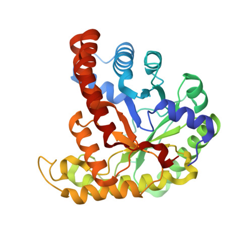 | |
UniProt | |||||
Entity Groups | |||||
| Sequence Clusters | 30% Identity50% Identity70% Identity90% Identity95% Identity100% Identity | ||||
| UniProt Group | P63705 | ||||
Sequence AnnotationsExpand | |||||
Reference Sequence | |||||
| Ligands 3 Unique | |||||
|---|---|---|---|---|---|
| ID | Chains | Name / Formula / InChI Key | 2D Diagram | 3D Interactions | |
| G3P Download:Ideal Coordinates CCD File | E [auth A], J [auth B], L [auth C], O [auth D] | SN-GLYCEROL-3-PHOSPHATE C3 H9 O6 P AWUCVROLDVIAJX-GSVOUGTGSA-N |  | ||
| G3H Download:Ideal Coordinates CCD File | F [auth A], M [auth C] | GLYCERALDEHYDE-3-PHOSPHATE C3 H7 O6 P LXJXRIRHZLFYRP-VKHMYHEASA-N |  | ||
| CA Download:Ideal Coordinates CCD File | G [auth A] H [auth A] I [auth A] K [auth B] N [auth C] | CALCIUM ION Ca BHPQYMZQTOCNFJ-UHFFFAOYSA-N |  | ||
| Length ( Å ) | Angle ( ˚ ) |
|---|---|
| a = 64.081 | α = 90 |
| b = 108.19 | β = 90 |
| c = 238.04 | γ = 90 |
| Software Name | Purpose |
|---|---|
| PHENIX | refinement |
| HKL-2000 | data reduction |
| HKL-2000 | data scaling |
| PHENIX | phasing |


