5FM0 | pdb_00005fm0

Crystal structure of the 6-carboxyhexanoate-CoA ligase (BioW)from Bacillus subtilis (PtCl4 derivative)


Experimental Data Snapshot

  • Method: X-RAY DIFFRACTION
  • Resolution: 2.44 Å
  • R-Value Free: 
    0.242 (Depositor) 
  • R-Value Work: 
    0.198 (Depositor) 
  • R-Value Observed: 
    0.201 (Depositor) 

wwPDB Validation 3D Report Full Report

Validation slider image for 5FM0

Ligand Structure Quality Assessment 


This is version 1.4 of the entry. See complete history

Literature

Using the pimeloyl-CoA synthetase adenylation fold to synthesize fatty acid thioesters.

Wang, M.Moynie, L.Harrison, P.J.Kelly, V.Piper, A.Naismith, J.H.Campopiano, D.J.

(2017) Nat Chem Biol 13: 660-667

  • DOI: https://doi.org/10.1038/nchembio.2361
  • Primary Citation Related Structures: 
    5FLG, 5FLL, 5FM0

  • PubMed Abstract: 

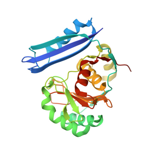
    Biotin is an essential vitamin in plants and mammals, functioning as the carbon dioxide carrier within central lipid metabolism. Bacterial pimeloyl-CoA synthetase (BioW) acts as a highly specific substrate-selection gate, ensuring the integrity of the carbon chain in biotin synthesis. BioW catalyzes the condensation of pimelic acid (C7 dicarboxylic acid) with CoASH in an ATP-dependent manner to form pimeloyl-CoA, the first dedicated biotin building block. Multiple structures of Bacillus subtilis BioW together capture all three substrates, as well as the intermediate pimeloyl-adenylate and product pyrophosphate (PP i ), indicating that the enzyme uses an internal ruler to select the correct dicarboxylic acid substrate. Both the catalytic mechanism and the surprising stability of the adenylate intermediate were rationalized through site-directed mutagenesis. Building on this understanding, BioW was engineered to synthesize high-value heptanoyl (C7) and octanoyl (C8) monocarboxylic acid-CoA and C8 dicarboxylic-CoA products, highlighting the enzyme's synthetic potential.


  • Organizational Affiliation
    • EastChem School of Chemistry, University of Edinburgh, Edinburgh, UK.

Macromolecule Content 

  • Total Structure Weight: 61.9 kDa 
  • Atom Count: 4,097 
  • Modeled Residue Count: 494 
  • Deposited Residue Count: 520 
  • Unique protein chains: 1

Macromolecules

Find similar proteins by:|  3D Structure
Entity ID: 1
MoleculeChains  Sequence LengthOrganismDetailsImage
6-CARBOXYHEXANOATE--COA LIGASE
A, B
260Bacillus subtilisMutation(s): 0 
EC: 6.2.1.14
UniProt
Find proteins for P53559 (Bacillus subtilis (strain 168))
Explore P53559 
Go to UniProtKB:  P53559
Entity Groups
Sequence Clusters30% Identity50% Identity70% Identity90% Identity95% Identity100% Identity
UniProt GroupP53559
Sequence Annotations
Expand
Reference Sequence

Small Molecules

Ligands 4 Unique
IDChains Name / Formula / InChI Key2D Diagram3D Interactions
WAQ

Query on WAQ


Download:Ideal Coordinates CCD File
C [auth A],
J [auth B]
PIMELOYL-AMP
C17 H24 N5 O10 P
CPQFOEZUJPOPAM-KRQFVHPKSA-N
PT

Query on PT


Download:Ideal Coordinates CCD File
G [auth A]
H [auth A]
I [auth A]
L [auth B]
M [auth B]
G [auth A],
H [auth A],
I [auth A],
L [auth B],
M [auth B],
N [auth B]
PLATINUM (II) ION
Pt
HRGDZIGMBDGFTC-UHFFFAOYSA-N
PPV

Query on PPV


Download:Ideal Coordinates CCD File
D [auth A],
K [auth B]
PYROPHOSPHATE
H4 O7 P2
XPPKVPWEQAFLFU-UHFFFAOYSA-N
MG

Query on MG


Download:Ideal Coordinates CCD File
E [auth A],
F [auth A]
MAGNESIUM ION
Mg
JLVVSXFLKOJNIY-UHFFFAOYSA-N

Experimental Data & Validation

Experimental Data

  • Method: X-RAY DIFFRACTION
  • Resolution: 2.44 Å
  • R-Value Free:  0.242 (Depositor) 
  • R-Value Work:  0.198 (Depositor) 
  • R-Value Observed: 0.201 (Depositor) 
Space Group: P 21 21 21
Unit Cell:
Length ( Å )Angle ( ˚ )
a = 50.61α = 90
b = 78.36β = 90
c = 166.7γ = 90
Software Package:
Software NamePurpose
REFMACrefinement
XDSdata reduction
SCALAdata scaling
SHELXphasing

Structure Validation

View Full Validation Report


Ligand Structure Quality Assessment 


Entry History 

Deposition Data

Revision History  (Full details and data files)

  • Version 1.0: 2016-11-16
    Type: Initial release
  • Version 1.1: 2017-04-12
    Changes: Database references
  • Version 1.2: 2017-04-26
    Changes: Database references
  • Version 1.3: 2017-05-24
    Changes: Database references
  • Version 1.4: 2024-05-08
    Changes: Data collection, Database references, Derived calculations, Other, Refinement description