5FPH

The GTPase domains of the immunity-related Irga6 dimerize in a parallel head-to-head fashion


Experimental Data Snapshot

  • Method: X-RAY DIFFRACTION
  • Resolution: 3.20 Å
  • R-Value Free: 
    0.317 (Depositor), 0.320 (DCC) 
  • R-Value Work: 
    0.297 (Depositor), 0.300 (DCC) 
  • R-Value Observed: 
    0.298 (Depositor) 

Starting Model: experimental
View more details

wwPDB Validation   3D Report Full Report


Ligand Structure Quality Assessment 

Created with Raphaël 2.3.0Worse 01 BetterLigand structure goodness of fit to experimental dataBest fitted GNPClick on this verticalbar to view details

This is version 2.2 of the entry. See complete history


Literature

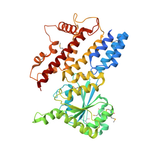
The Immunity-Related Gtpase Irga6 Dimerizes in a Parallel Head-to-Head Fashion.

Schulte, K.Pawlowski, N.Faelber, K.Froehlich, C.Howard, J.Daumke, O.

(2016) BMC Biol 14: 14

  • DOI: https://doi.org/10.1186/s12915-016-0236-7
  • Primary Citation of Related Structures:  
    5FPH

  • PubMed Abstract: 

    The immunity-related GTPases (IRGs) constitute a powerful cell-autonomous resistance system against several intracellular pathogens. Irga6 is a dynamin-like protein that oligomerizes at the parasitophorous vacuolar membrane (PVM) of Toxoplasma gondii leading to its vesiculation. Based on a previous biochemical analysis, it has been proposed that the GTPase domains of Irga6 dimerize in an antiparallel fashion during oligomerization. We determined the crystal structure of an oligomerization-impaired Irga6 mutant bound to a non-hydrolyzable GTP analog. Contrary to the previous model, the structure shows that the GTPase domains dimerize in a parallel fashion. The nucleotides in the center of the interface participate in dimerization by forming symmetric contacts with each other and with the switch I region of the opposing Irga6 molecule. The latter contact appears to activate GTP hydrolysis by stabilizing the position of the catalytic glutamate 106 in switch I close to the active site. Further dimerization contacts involve switch II, the G4 helix and the trans stabilizing loop. The Irga6 structure features a parallel GTPase domain dimer, which appears to be a unifying feature of all dynamin and septin superfamily members. This study contributes important insights into the assembly and catalytic mechanisms of IRG proteins as prerequisite to understand their anti-microbial action.


  • Organizational Affiliation

    Max-Delbrueck-Centrum for Molecular Medicine, Crystallography, Robert-Rössle-Strasse 10, 13125, Berlin, Germany.


Macromolecules
Find similar proteins by:  (by identity cutoff)  |  3D Structure
Entity ID: 1
MoleculeChains Sequence LengthOrganismDetailsImage
INTERFERON-INDUCIBLE GTPASE 1
A, B, C, D, E
423Mus musculusMutation(s): 4 
EC: 3.6.5
UniProt
Find proteins for Q9QZ85 (Mus musculus)
Explore Q9QZ85 
Go to UniProtKB:  Q9QZ85
Entity Groups  
Sequence Clusters30% Identity50% Identity70% Identity90% Identity95% Identity100% Identity
UniProt GroupQ9QZ85
Sequence Annotations
Expand
  • Reference Sequence
Small Molecules
Ligands 3 Unique
IDChains Name / Formula / InChI Key2D Diagram3D Interactions
GNP
Query on GNP

Download Ideal Coordinates CCD File 
H [auth A]
K [auth B]
N [auth C]
S [auth E]
W [auth F]
PHOSPHOAMINOPHOSPHONIC ACID-GUANYLATE ESTER
C10 H17 N6 O13 P3
UQABYHGXWYXDTK-UUOKFMHZSA-N
TRS
Query on TRS

Download Ideal Coordinates CCD File 
Q [auth C],
V [auth E]
2-AMINO-2-HYDROXYMETHYL-PROPANE-1,3-DIOL
C4 H12 N O3
LENZDBCJOHFCAS-UHFFFAOYSA-O
MG
Query on MG

Download Ideal Coordinates CCD File 
AA [auth G]
BA [auth G]
I [auth A]
J [auth A]
L [auth B]
MAGNESIUM ION
Mg
JLVVSXFLKOJNIY-UHFFFAOYSA-N
Modified Residues  1 Unique
IDChains TypeFormula2D DiagramParent
MSE
Query on MSE
A, B, C, D, E
L-PEPTIDE LINKINGC5 H11 N O2 SeMET
Experimental Data & Validation

Experimental Data

  • Method: X-RAY DIFFRACTION
  • Resolution: 3.20 Å
  • R-Value Free:  0.317 (Depositor), 0.320 (DCC) 
  • R-Value Work:  0.297 (Depositor), 0.300 (DCC) 
  • R-Value Observed: 0.298 (Depositor) 
Space Group: P 61 2 2
Unit Cell:
Length ( Å )Angle ( ˚ )
a = 98.51α = 90
b = 98.51β = 90
c = 1289.4γ = 120
Software Package:
Software NamePurpose
PHENIXrefinement
XDSdata reduction
XSCALEdata scaling
PHENIXphasing

Structure Validation

View Full Validation Report


Ligand Structure Quality Assessment 

Created with Raphaël 2.3.0Worse 01 BetterLigand structure goodness of fit to experimental dataBest fitted GNPClick on this verticalbar to view details

Entry History 

Deposition Data

Revision History  (Full details and data files)

  • Version 1.0: 2016-02-10
    Type: Initial release
  • Version 1.1: 2016-03-16
    Changes: Database references
  • Version 1.2: 2017-07-12
    Changes: Derived calculations
  • Version 2.0: 2021-08-11
    Changes: Advisory, Atomic model, Data collection, Database references, Derived calculations, Other
  • Version 2.1: 2024-01-10
    Changes: Data collection, Refinement description
  • Version 2.2: 2024-11-06
    Changes: Structure summary