5FX3

Breaking down the wall: mutation of the tyrosine gate of the universal Escherichia coli fimbrial adhesin FimH


Experimental Data Snapshot

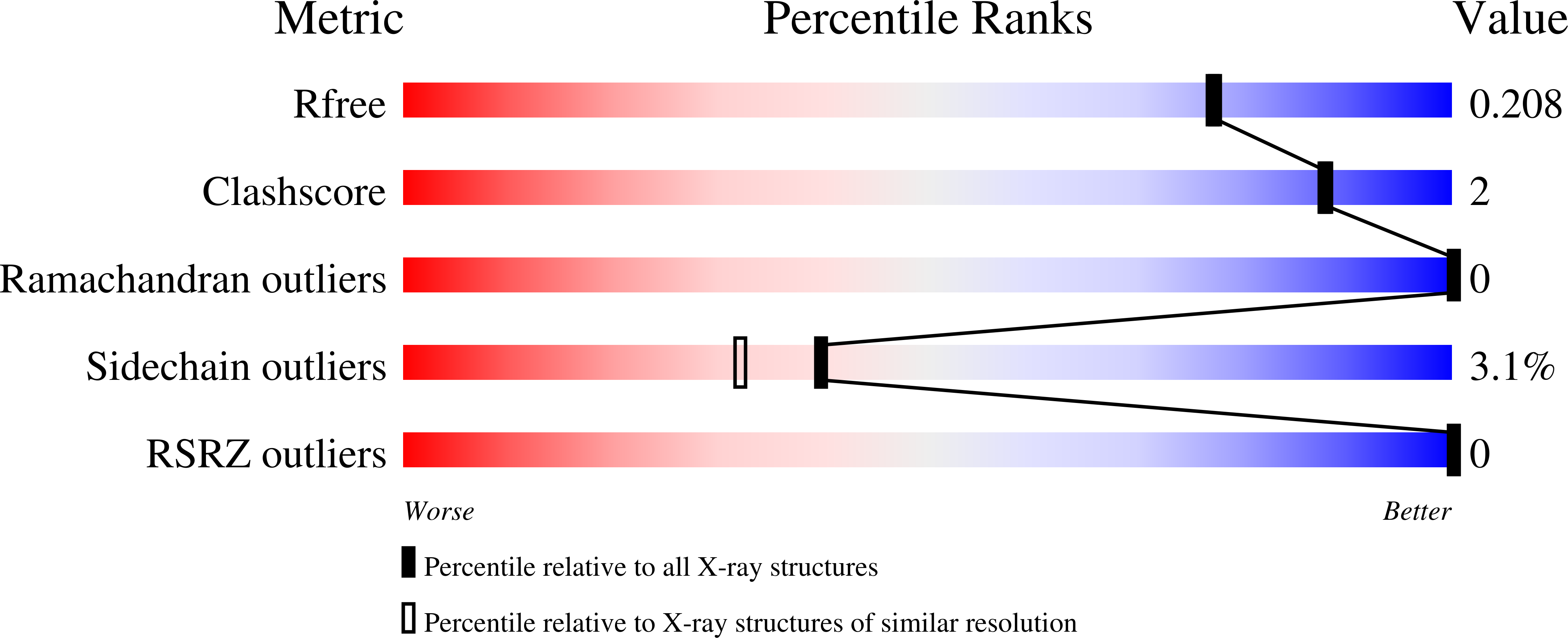
  • Method: X-RAY DIFFRACTION
  • Resolution: 1.90 Å
  • R-Value Free: 0.202 
  • R-Value Work: 0.158 
  • R-Value Observed: 0.161 

Starting Model: experimental
View more details

wwPDB Validation   3D Report Full Report


Ligand Structure Quality Assessment 


This is version 2.2 of the entry. See complete history


Literature

Mutation of Tyr137 of the universal Escherichia coli fimbrial adhesin FimH relaxes the tyrosine gate prior to mannose binding.

Rabbani, S.Krammer, E.M.Roos, G.Zalewski, A.Preston, R.Eid, S.Zihlmann, P.Prevost, M.Lensink, M.F.Thompson, A.Ernst, B.Bouckaert, J.

(2017) IUCrJ 4: 7-23

  • DOI: https://doi.org/10.1107/S2052252516016675
  • Primary Citation of Related Structures:  
    4CA4, 5FS5, 5FWR, 5FX3

  • PubMed Abstract: 

    The most prevalent diseases manifested by Escherichia coli are acute and recurrent bladder infections and chronic inflammatory bowel diseases such as Crohn's disease. E. coli clinical isolates express the FimH adhesin, which consists of a mannose-specific lectin domain connected via a pilin domain to the tip of type 1 pili. Although the isolated FimH lectin domain has affinities in the nanomolar range for all high-mannosidic glycans, differentiation between these glycans is based on their capacity to form predominantly hydrophobic interactions within the tyrosine gate at the entrance to the binding pocket. In this study, novel crystal structures of tyrosine-gate mutants of FimH, ligand-free or in complex with heptyl α-d- O -mannopyranoside or 4-biphenyl α-d- O- mannopyranoside, are combined with quantum-mechanical calculations and molecular-dynamics simulations. In the Y48A FimH crystal structure, a large increase in the dynamics of the alkyl chain of heptyl α-d- O -mannopyranoside attempts to compensate for the absence of the aromatic ring; however, the highly energetic and stringent mannose-binding pocket of wild-type FimH is largely maintained. The Y137A mutation, on the other hand, is the most detrimental to FimH affinity and specificity: (i) in the absence of ligand the FimH C-terminal residue Thr158 intrudes into the mannose-binding pocket and (ii) ethylenediaminetetraacetic acid interacts strongly with Glu50, Thr53 and Asn136, in spite of multiple dialysis and purification steps. Upon mutation, pre-ligand-binding relaxation of the backbone dihedral angles at position 137 in the tyrosine gate and their coupling to Tyr48 via the interiorly located Ile52 form the basis of the loss of affinity of the FimH adhesin in the Y137A mutant.


  • Organizational Affiliation

    Institute of Molecular Pharmacy, Pharmacenter, University of Basel , Klingelbergstrasse 50-70, CH-4056 Basel, Switzerland.


Macromolecules
Find similar proteins by:  (by identity cutoff)  |  3D Structure
Entity ID: 1
MoleculeChains Sequence LengthOrganismDetailsImage
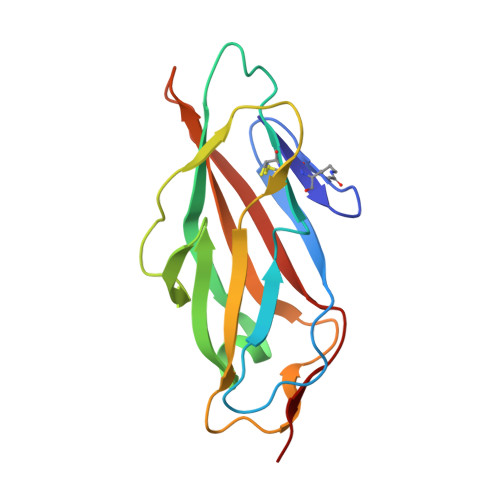
FimH158Escherichia coliMutation(s): 1 
Gene Names: fimH
UniProt
Find proteins for P08191 (Escherichia coli (strain K12))
Explore P08191 
Go to UniProtKB:  P08191
Entity Groups  
Sequence Clusters30% Identity50% Identity70% Identity90% Identity95% Identity100% Identity
UniProt GroupP08191
Sequence Annotations
Expand
  • Reference Sequence
Small Molecules
Ligands 1 Unique
IDChains Name / Formula / InChI Key2D Diagram3D Interactions
EDT
Query on EDT

Download Ideal Coordinates CCD File 
B [auth A]{[-(BIS-CARBOXYMETHYL-AMINO)-ETHYL]-CARBOXYMETHYL-AMINO}-ACETIC ACID
C10 H16 N2 O8
KCXVZYZYPLLWCC-UHFFFAOYSA-N
Modified Residues  1 Unique
IDChains TypeFormula2D DiagramParent
ALY
Query on ALY
A
L-PEPTIDE LINKINGC8 H16 N2 O3LYS
Experimental Data & Validation

Experimental Data

  • Method: X-RAY DIFFRACTION
  • Resolution: 1.90 Å
  • R-Value Free: 0.202 
  • R-Value Work: 0.158 
  • R-Value Observed: 0.161 
  • Space Group: P 65 2 2
Unit Cell:
Length ( Å )Angle ( ˚ )
a = 54.18α = 90
b = 54.18β = 90
c = 257.95γ = 120
Software Package:
Software NamePurpose
PHENIXrefinement
XDSdata reduction
XDSdata scaling
PHASERphasing

Structure Validation

View Full Validation Report


Ligand Structure Quality Assessment 


Entry History 

Deposition Data

Revision History  (Full details and data files)

  • Version 1.0: 2017-01-18
    Type: Initial release
  • Version 1.1: 2017-03-15
    Changes: Database references
  • Version 1.2: 2017-09-13
    Changes: Data collection
  • Version 1.3: 2019-09-25
    Changes: Data collection, Database references, Derived calculations, Other, Source and taxonomy, Structure summary
  • Version 1.4: 2023-09-27
    Changes: Data collection, Database references, Derived calculations
  • Version 2.0: 2023-11-15
    Changes: Atomic model, Data collection
  • Version 2.1: 2024-01-10
    Changes: Refinement description
  • Version 2.2: 2024-10-16
    Changes: Structure summary