


Crystal structure of a bacterial fucosidase with iminocyclitol (2S,3S,4R,5S)-3,4-dihydroxy-2-ethynyl-5-methylpyrrolidine
Wright, D.W., Davies, G.J.To be published.
Experimental Data Snapshot
Starting Model: experimental
View more details
Entity ID: 1 | |||||
|---|---|---|---|---|---|
| Molecule | Chains | Sequence Length | Organism | Details | Image |
| Alpha-L-fucosidase | 447 | Bacteroides thetaiotaomicron VPI-5482 | Mutation(s): 0 Gene Names: BT_2970 EC: 3.2.1.51 | 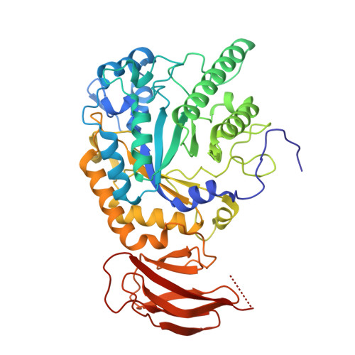 | |
UniProt | |||||
Entity Groups | |||||
| Sequence Clusters | 30% Identity50% Identity70% Identity90% Identity95% Identity100% Identity | ||||
| UniProt Group | Q8A3I4 | ||||
Sequence AnnotationsExpand | |||||
Reference Sequence | |||||
| Ligands 3 Unique | |||||
|---|---|---|---|---|---|
| ID | Chains | Name / Formula / InChI Key | 2D Diagram | 3D Interactions | |
| H79 Download:Ideal Coordinates CCD File | H [auth A], L [auth B], O [auth C], Q [auth D] | [(1,2,3,4,5-eta)-cyclopentadienyl][(1,2,3,4,5-eta)-1-{3-[4-(3,4-dihydroxy-5-methylpyrrolidin-2-yl)-1H-1,2,3-triazol-1-yl]prop-1-en-1-yl}cyclopentadienyl]iron C20 H15 Fe N4 O2 FQODPMWJAJVXMD-YCHRPOLYSA-N |  | ||
| SO4 Download:Ideal Coordinates CCD File | E [auth A], F [auth A], I [auth B], J [auth B], M [auth C] | SULFATE ION O4 S QAOWNCQODCNURD-UHFFFAOYSA-L |  | ||
| IMD Download:Ideal Coordinates CCD File | G [auth A], K [auth B], N [auth C], P [auth D] | IMIDAZOLE C3 H5 N2 RAXXELZNTBOGNW-UHFFFAOYSA-O |  | ||
| Length ( Å ) | Angle ( ˚ ) |
|---|---|
| a = 56.5 | α = 90 |
| b = 188.76 | β = 93.89 |
| c = 98.14 | γ = 90 |
| Software Name | Purpose |
|---|---|
| REFMAC | refinement |
| PDB_EXTRACT | data extraction |
| Aimless | data scaling |
| Funding Organization | Location | Grant Number |
|---|---|---|
| Biotechnology and Biological Sciences Research Council | United Kingdom | -- |


