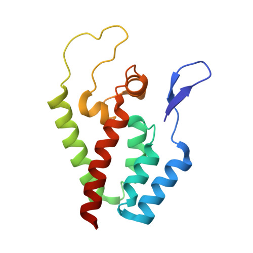
5HGK

HIV-1 CA N-terminal domain, open conformation


Experimental Data Snapshot

  • Method: X-RAY DIFFRACTION
  • Resolution: 1.76 Å
  • R-Value Free: 0.224 
  • R-Value Work: 0.190 
  • R-Value Observed: 0.192 

Starting Model: experimental
View more details

wwPDB Validation   3D Report Full Report


This is version 1.5 of the entry. See complete history


Literature

HIV-1 uses dynamic capsid pores to import nucleotides and fuel encapsidated DNA synthesis.

Jacques, D.A.McEwan, W.A.Hilditch, L.Price, A.J.Towers, G.J.James, L.C.

(2016) Nature 536: 349-353

  • DOI: https://doi.org/10.1038/nature19098
  • Primary Citation of Related Structures:  
    5HGK, 5HGL, 5HGM, 5HGN, 5HGO, 5HGP, 5JPA

  • PubMed Abstract: 

    During the early stages of infection, the HIV-1 capsid protects viral components from cytosolic sensors and nucleases such as cGAS and TREX, respectively, while allowing access to nucleotides for efficient reverse transcription. Here we show that each capsid hexamer has a size-selective pore bound by a ring of six arginine residues and a 'molecular iris' formed by the amino-terminal β-hairpin. The arginine ring creates a strongly positively charged channel that recruits the four nucleotides with on-rates that approach diffusion limits. Progressive removal of pore arginines results in a dose-dependent and concomitant decrease in nucleotide affinity, reverse transcription and infectivity. This positively charged channel is universally conserved in lentiviral capsids despite the fact that it is strongly destabilizing without nucleotides to counteract charge repulsion. We also describe a channel inhibitor, hexacarboxybenzene, which competes for nucleotide binding and efficiently blocks encapsidated reverse transcription, demonstrating the tractability of the pore as a novel drug target.


Macromolecules
Find similar proteins by:  (by identity cutoff)  |  3D Structure
Entity ID: 1
MoleculeChains Sequence LengthOrganismDetailsImage
Capsid protein
A, B
146Human immunodeficiency virus 1Mutation(s): 0 
UniProt
Find proteins for A9PKC6 (Human immunodeficiency virus type 1)
Explore A9PKC6 
Go to UniProtKB:  A9PKC6
Entity Groups  
Sequence Clusters30% Identity50% Identity70% Identity90% Identity95% Identity100% Identity
UniProt GroupA9PKC6
Sequence Annotations
Expand
  • Reference Sequence
Small Molecules
Ligands 1 Unique
IDChains Name / Formula / InChI Key2D Diagram3D Interactions
CL
Query on CL

Download Ideal Coordinates CCD File 
C [auth B]CHLORIDE ION
Cl
VEXZGXHMUGYJMC-UHFFFAOYSA-M
Experimental Data & Validation

Experimental Data

  • Method: X-RAY DIFFRACTION
  • Resolution: 1.76 Å
  • R-Value Free: 0.224 
  • R-Value Work: 0.190 
  • R-Value Observed: 0.192 
  • Space Group: P 1 21 1
Unit Cell:
Length ( Å )Angle ( ˚ )
a = 43.72α = 90
b = 23.85β = 96.31
c = 129.549γ = 90
Software Package:
Software NamePurpose
MAR345dtbdata collection
SCALAdata scaling
PHASERphasing
REFMACrefinement
PDB_EXTRACTdata extraction
iMOSFLMdata reduction

Structure Validation

View Full Validation Report


Entry History & Funding Information

Deposition Data


Funding OrganizationLocationGrant Number
Medical Research Council (United Kingdom)United KingdomUK; U10518101
European Research CouncilUnited Kingdom281627 -IAI
National Health and Medical Research CouncilAustraliaGNT1036521

Revision History  (Full details and data files)

  • Version 1.0: 2016-08-10
    Type: Initial release
  • Version 1.1: 2016-08-24
    Changes: Database references
  • Version 1.2: 2016-08-31
    Changes: Database references
  • Version 1.3: 2017-09-13
    Changes: Author supporting evidence
  • Version 1.4: 2018-01-24
    Changes: Source and taxonomy
  • Version 1.5: 2024-01-10
    Changes: Data collection, Database references, Refinement description