5HHT | pdb_00005hht

Crystal structure of E. coli transketolase triple variant Ser385Tyr/Asp469Thr/Arg520Gln


Experimental Data Snapshot

  • Method: X-RAY DIFFRACTION
  • Resolution: 1.50 Å
  • R-Value Free: 
    0.157 (Depositor), 0.157 (DCC) 
  • R-Value Work: 
    0.126 (Depositor), 0.127 (DCC) 
  • R-Value Observed: 
    0.127 (Depositor) 

wwPDB Validation 3D Report Full Report

Validation slider image for 5HHT

Ligand Structure Quality Assessment 


This is version 2.1 of the entry. See complete history

Literature

Structural Analysis of an Evolved Transketolase Reveals Divergent Binding Modes.

Affaticati, P.E.Dai, S.B.Payongsri, P.Hailes, H.C.Tittmann, K.Dalby, P.A.

(2016) Sci Rep 6: 35716-35716

  • DOI: https://doi.org/10.1038/srep35716
  • Primary Citation Related Structures: 
    5HHT

  • PubMed Abstract: 

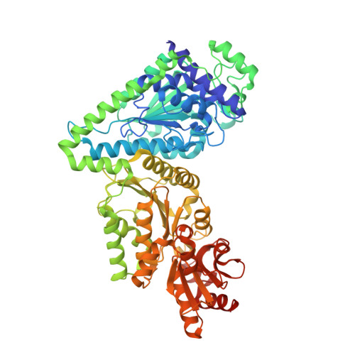
    The S385Y/D469T/R520Q variant of E. coli transketolase was evolved previously with three successive smart libraries, each guided by different structural, bioinformatical or computational methods. Substrate-walking progressively shifted the target acceptor substrate from phosphorylated aldehydes, towards a non-phosphorylated polar aldehyde, a non-polar aliphatic aldehyde, and finally a non-polar aromatic aldehyde. Kinetic evaluations on three benzaldehyde derivatives, suggested that their active-site binding was differentially sensitive to the S385Y mutation. Docking into mutants generated in silico from the wild-type crystal structure was not wholly satisfactory, as errors accumulated with successive mutations, and hampered further smart-library designs. Here we report the crystal structure of the S385Y/D469T/R520Q variant, and molecular docking of three substrates. This now supports our original hypothesis that directed-evolution had generated an evolutionary intermediate with divergent binding modes for the three aromatic aldehydes tested. The new active site contained two binding pockets supporting π-π stacking interactions, sterically separated by the D469T mutation. While 3-formylbenzoic acid (3-FBA) preferred one pocket, and 4-FBA the other, the less well-accepted substrate 3-hydroxybenzaldehyde (3-HBA) was caught in limbo with equal preference for the two pockets. This work highlights the value of obtaining crystal structures of evolved enzyme variants, for continued and reliable use of smart library strategies.


  • Organizational Affiliation
    • Department of Biochemical Engineering, Gordon Street, University College London, WC1H 0AH, UK.

Macromolecule Content 

  • Total Structure Weight: 149.47 kDa 
  • Atom Count: 12,313 
  • Modeled Residue Count: 1,329 
  • Deposited Residue Count: 1,338 
  • Unique protein chains: 1

Macromolecules

Find similar proteins by:|  3D Structure
Entity ID: 1
MoleculeChains  Sequence LengthOrganismDetailsImage
Transketolase 1
A, B
669Escherichia coli K-12Mutation(s): 3 
Gene Names: tktAtktb2935JW5478
EC: 2.2.1.1
UniProt
Find proteins for P27302 (Escherichia coli (strain K12))
Explore P27302 
Go to UniProtKB:  P27302
Entity Groups
Sequence Clusters30% Identity50% Identity70% Identity90% Identity95% Identity100% Identity
UniProt GroupP27302
Sequence Annotations
Expand
Reference Sequence

Small Molecules

Ligands 3 Unique
IDChains Name / Formula / InChI Key2D Diagram3D Interactions
TPP

Query on TPP


Download:Ideal Coordinates CCD File
DA [auth B],
E [auth A]
THIAMINE DIPHOSPHATE
C12 H19 N4 O7 P2 S
AYEKOFBPNLCAJY-UHFFFAOYSA-O
EDO

Query on EDO


Download:Ideal Coordinates CCD File
AA [auth B]
BA [auth B]
C [auth A]
CA [auth B]
F [auth A]
AA [auth B],
BA [auth B],
C [auth A],
CA [auth B],
F [auth A],
FA [auth B],
G [auth A],
GA [auth B],
H [auth A],
HA [auth B],
I [auth A],
IA [auth B],
J [auth A],
JA [auth B],
K [auth A],
KA [auth B],
L [auth A],
LA [auth B],
M [auth A],
MA [auth B],
N [auth A],
NA [auth B],
O [auth A],
OA [auth B],
P [auth A],
PA [auth B],
Q [auth A],
R [auth A],
S [auth A],
T [auth B],
U [auth B],
V [auth B],
W [auth B],
X [auth B],
Y [auth B],
Z [auth B]
1,2-ETHANEDIOL
C2 H6 O2
LYCAIKOWRPUZTN-UHFFFAOYSA-N
CA

Query on CA


Download:Ideal Coordinates CCD File
D [auth A],
EA [auth B]
CALCIUM ION
Ca
BHPQYMZQTOCNFJ-UHFFFAOYSA-N

Experimental Data & Validation

Experimental Data

  • Method: X-RAY DIFFRACTION
  • Resolution: 1.50 Å
  • R-Value Free:  0.157 (Depositor), 0.157 (DCC) 
  • R-Value Work:  0.126 (Depositor), 0.127 (DCC) 
  • R-Value Observed: 0.127 (Depositor) 
Space Group: P 21 21 21
Unit Cell:
Length ( Å )Angle ( ˚ )
a = 89.944α = 90
b = 102.044β = 90
c = 133.076γ = 90
Software Package:
Software NamePurpose
PHENIXrefinement
XSCALEdata scaling
PDB_EXTRACTdata extraction
XDSdata reduction
PHENIXphasing

Structure Validation

View Full Validation Report


Ligand Structure Quality Assessment 


Entry History 

& Funding Information

Deposition Data


Funding OrganizationLocationGrant Number
German Research FoundationGermanyFOR 1296/TP 3

Revision History  (Full details and data files)

  • Version 1.0: 2016-11-02
    Type: Initial release
  • Version 1.1: 2017-09-06
    Changes: Author supporting evidence, Data collection
  • Version 2.0: 2021-08-04
    Changes: Atomic model, Data collection, Derived calculations, Non-polymer description, Structure summary
  • Version 2.1: 2024-05-08
    Changes: Data collection, Database references