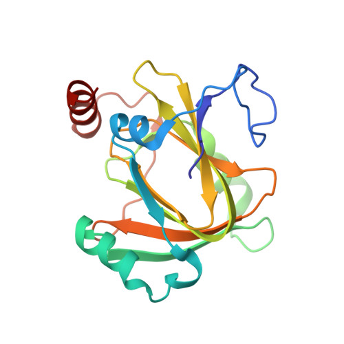
5I8Y

Structure of Mouse Acireductone Dioxygenase bound to Co2+ and 2-keto-4-(methylthio)-butyric acid


Experimental Data Snapshot

  • Method: X-RAY DIFFRACTION
  • Resolution: 1.94 Å
  • R-Value Free: 0.191 
  • R-Value Work: 0.159 
  • R-Value Observed: 0.162 

Starting Model: experimental
View more details

wwPDB Validation   3D Report Full Report


This is version 1.5 of the entry. See complete history


Literature

Metal-Dependent Function of a Mammalian Acireductone Dioxygenase.

Deshpande, A.R.Wagenpfeil, K.Pochapsky, T.C.Petsko, G.A.Ringe, D.

(2016) Biochemistry 55: 1398-1407

  • DOI: https://doi.org/10.1021/acs.biochem.5b01319
  • Primary Citation of Related Structures:  
    5I8S, 5I8T, 5I8Y, 5I91, 5I93

  • PubMed Abstract: 

    The two acireductone dioxygenase (ARD) isozymes from the methionine salvage pathway of Klebsiella oxytoca are the only known pair of naturally occurring metalloenzymes with distinct chemical and physical properties determined solely by the identity of the divalent transition metal ion (Fe(2+) or Ni(2+)) in the active site. We now show that this dual chemistry can also occur in mammals. ARD from Mus musculus (MmARD) was studied to relate the metal ion identity and three-dimensional structure to enzyme function. The iron-containing isozyme catalyzes the cleavage of 1,2-dihydroxy-3-keto-5-(thiomethyl)pent-1-ene (acireductone) by O2 to formate and the ketoacid precursor of methionine, which is the penultimate step in methionine salvage. The nickel-bound form of ARD catalyzes an off-pathway reaction resulting in formate, carbon monoxide (CO), and 3-(thiomethyl) propionate. Recombinant MmARD was expressed and purified to obtain a homogeneous enzyme with a single transition metal ion bound. The Fe(2+)-bound protein, which shows about 10-fold higher activity than that of others, catalyzes on-pathway chemistry, whereas the Ni(2+), Co(2+), or Mn(2+) forms exhibit off-pathway chemistry, as has been seen with ARD from Klebsiella. Thermal stability of the isozymes is strongly affected by the metal ion identity, with Ni(2+)-bound MmARD being the most stable, followed by Co(2+) and Fe(2+), and Mn(2+)-bound ARD being the least stable. Ni(2+)- and Co(2+)-bound MmARD were crystallized, and the structures of the two proteins found to be similar. Enzyme-ligand complexes provide insight into substrate binding, metal coordination, and the catalytic mechanism.


  • Organizational Affiliation

    Helen and Robert Appel Alzheimer's Disease Research Institute, Weill Cornell Medical College , New York, New York 10065, United States.


Macromolecules
Find similar proteins by:  (by identity cutoff)  |  3D Structure
Entity ID: 1
MoleculeChains Sequence LengthOrganismDetailsImage
1,2-dihydroxy-3-keto-5-methylthiopentene dioxygenase179Mus musculusMutation(s): 0 
Gene Names: Adi1Mtcbp1
EC: 1.13.11.54
UniProt
Find proteins for Q99JT9 (Mus musculus)
Go to UniProtKB:  Q99JT9
Sequence Annotations
Expand
  • Reference Sequence
Small Molecules
Ligands 2 Unique
IDChains Name / Formula / InChI Key2D Diagram3D Interactions
KMT
Query on KMT

Download Ideal Coordinates CCD File 
C [auth A]4-(METHYLSULFANYL)-2-OXOBUTANOIC ACID
C5 H8 O3 S
SXFSQZDSUWACKX-UHFFFAOYSA-N
CO
Query on CO

Download Ideal Coordinates CCD File 
B [auth A]COBALT (II) ION
Co
XLJKHNWPARRRJB-UHFFFAOYSA-N
Experimental Data & Validation

Experimental Data

  • Method: X-RAY DIFFRACTION
  • Resolution: 1.94 Å
  • R-Value Free: 0.191 
  • R-Value Work: 0.159 
  • R-Value Observed: 0.162 
  • Space Group: P 41 21 2
Unit Cell:
Length ( Å )Angle ( ˚ )
a = 79.089α = 90
b = 79.089β = 90
c = 115.125γ = 90
Software Package:
Software NamePurpose
SCALEPACKdata scaling
PHASERphasing
PHENIXrefinement
PDB_EXTRACTdata extraction

Structure Validation

View Full Validation Report


Entry History & Funding Information

Deposition Data


Funding OrganizationLocationGrant Number
National Institutes of Health/National Institute of General Medical Sciences (NIH/NIGMS)United StatesGM26788

Revision History  (Full details and data files)

  • Version 1.0: 2016-03-09
    Type: Initial release
  • Version 1.1: 2016-03-23
    Changes: Database references
  • Version 1.2: 2017-09-27
    Changes: Author supporting evidence, Database references, Derived calculations
  • Version 1.3: 2019-12-25
    Changes: Author supporting evidence
  • Version 1.4: 2020-09-02
    Changes: Structure summary
  • Version 1.5: 2023-09-27
    Changes: Data collection, Database references, Refinement description