5IWH | pdb_00005iwh

Structure of P. vulgaris HigB toxin delta H92


Experimental Data Snapshot

  • Method: X-RAY DIFFRACTION
  • Resolution: 1.10 Å
  • R-Value Free: 
    0.169 (Depositor), 0.169 (DCC) 
  • R-Value Work: 
    0.138 (Depositor), 0.139 (DCC) 
  • R-Value Observed: 
    0.140 (Depositor) 

Starting Model: experimental
View more details

wwPDB Validation 3D Report Full Report

Validation slider image for 5IWH

This is version 1.2 of the entry. See complete history

Literature

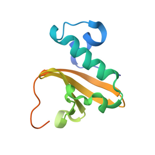
Mechanism of endonuclease cleavage by the HigB toxin.

Schureck, M.A.Repack, A.Miles, S.J.Marquez, J.Dunham, C.M.

(2016) Nucleic Acids Res 44: 7944-7953

  • DOI: https://doi.org/10.1093/nar/gkw598
  • Primary Citation Related Structures: 
    4ZSN, 5IWH, 5IXL

  • PubMed Abstract: 

    Bacteria encode multiple type II toxin-antitoxin modules that cleave ribosome-bound mRNAs in response to stress. All ribosome-dependent toxin family members structurally characterized to date adopt similar microbial RNase architectures despite possessing low sequence identities. Therefore, determining which residues are catalytically important in this specialized RNase family has been a challenge in the field. Structural studies of RelE and YoeB toxins bound to the ribosome provided significant insights but biochemical experiments with RelE were required to clearly demonstrate which residues are critical for acid-base catalysis of mRNA cleavage. Here, we solved an X-ray crystal structure of the wild-type, ribosome-dependent toxin HigB bound to the ribosome revealing potential catalytic residues proximal to the mRNA substrate. Using cell-based and biochemical assays, we further determined that HigB residues His54, Asp90, Tyr91 and His92 are critical for activity in vivo, while HigB H54A and Y91A variants have the largest effect on mRNA cleavage in vitro Comparison of X-ray crystal structures of two catalytically inactive HigB variants with 70S-HigB bound structures reveal that HigB active site residues undergo conformational rearrangements likely required for recognition of its mRNA substrate. These data support the emerging concept that ribosome-dependent toxins have diverse modes of mRNA recognition.


  • Organizational Affiliation
    • Emory University School of Medicine, Department of Biochemistry, 1510 Clifton Road NE, Atlanta, GA 30322, USA.

Macromolecule Content 

  • Total Structure Weight: 13.65 kDa 
  • Atom Count: 1,020 
  • Modeled Residue Count: 97 
  • Deposited Residue Count: 117 
  • Unique protein chains: 1

Macromolecules

Find similar proteins by:|  3D Structure
Entity ID: 1
MoleculeChains  Sequence LengthOrganismDetailsImage
Endoribonuclease HigB117Proteus vulgarisMutation(s): 0 
Gene Names: higB
EC: 3.1
UniProt
Find proteins for Q7A225 (Proteus vulgaris)
Explore Q7A225 
Go to UniProtKB:  Q7A225
Entity Groups
Sequence Clusters30% Identity50% Identity70% Identity90% Identity95% Identity100% Identity
UniProt GroupQ7A225
Sequence Annotations
Expand
Reference Sequence

Experimental Data & Validation

Experimental Data

  • Method: X-RAY DIFFRACTION
  • Resolution: 1.10 Å
  • R-Value Free:  0.169 (Depositor), 0.169 (DCC) 
  • R-Value Work:  0.138 (Depositor), 0.139 (DCC) 
  • R-Value Observed: 0.140 (Depositor) 
Space Group: C 1 2 1
Unit Cell:
Length ( Å )Angle ( ˚ )
a = 55.776α = 90
b = 33.166β = 115.49
c = 51.492γ = 90
Software Package:
Software NamePurpose
PHENIXrefinement
HKL-2000data reduction
HKL-2000data scaling
PHENIXphasing

Structure Validation

View Full Validation Report


Entry History 

Deposition Data

Revision History  (Full details and data files)

  • Version 1.0: 2016-07-20
    Type: Initial release
  • Version 1.1: 2016-10-05
    Changes: Database references
  • Version 1.2: 2023-09-27
    Changes: Data collection, Database references, Derived calculations, Refinement description