5IXQ | pdb_00005ixq

Crystal structure of the Arabidopsis receptor kinase HAESA LRR ectdomain in complex with the peptide hormone IDA.


Experimental Data Snapshot

  • Method: X-RAY DIFFRACTION
  • Resolution: 1.86 Å
  • R-Value Free: 
    0.207 (Depositor), 0.215 (DCC) 
  • R-Value Work: 
    0.183 (Depositor), 0.190 (DCC) 
  • R-Value Observed: 
    0.184 (Depositor) 

Starting Model: experimental
View more details

wwPDB Validation 3D Report Full Report

Validation slider image for 5IXQ

Ligand Structure Quality Assessment 


This is version 2.1 of the entry. See complete history

Literature

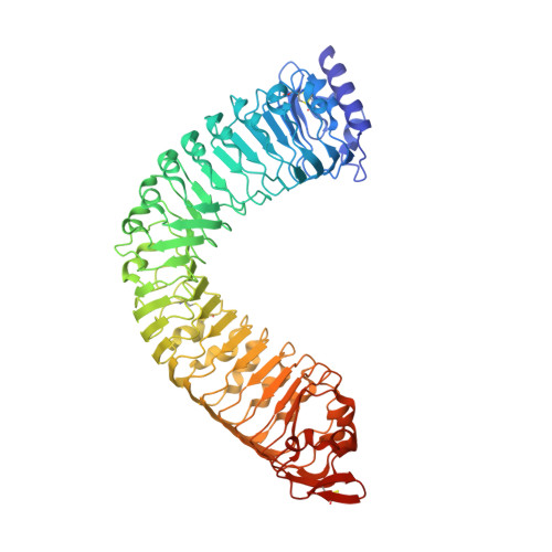
Mechanistic insight into a peptide hormone signaling complex mediating floral organ abscission.

Santiago, J.Brandt, B.Wildhagen, M.Hohmann, U.Hothorn, L.A.Butenko, M.A.Hothorn, M.

(2016) Elife 5

  • DOI: https://doi.org/10.7554/eLife.15075
  • Primary Citation Related Structures: 
    5IXO, 5IXQ, 5IXT, 5IYV, 5IYX

  • PubMed Abstract: 

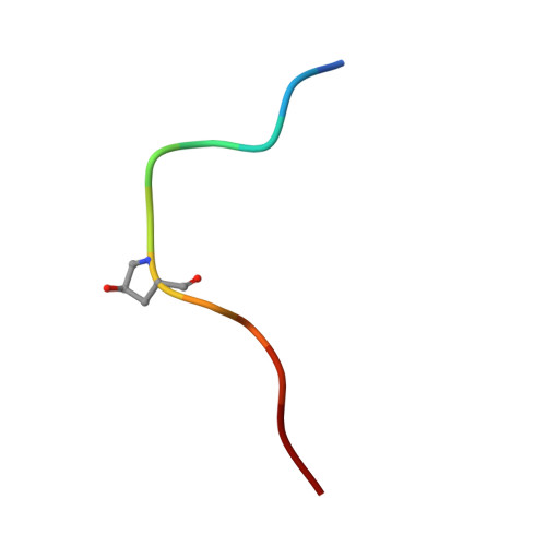
    Plants constantly renew during their life cycle and thus require to shed senescent and damaged organs. Floral abscission is controlled by the leucine-rich repeat receptor kinase (LRR-RK) HAESA and the peptide hormone IDA. It is unknown how expression of IDA in the abscission zone leads to HAESA activation. Here we show that IDA is sensed directly by the HAESA ectodomain. Crystal structures of HAESA in complex with IDA reveal a hormone binding pocket that accommodates an active dodecamer peptide. A central hydroxyproline residue anchors IDA to the receptor. The HAESA co-receptor SERK1, a positive regulator of the floral abscission pathway, allows for high-affinity sensing of the peptide hormone by binding to an Arg-His-Asn motif in IDA. This sequence pattern is conserved among diverse plant peptides, suggesting that plant peptide hormone receptors may share a common ligand binding mode and activation mechanism.


  • Organizational Affiliation
    • Structural Plant Biology Laboratory, Department of Botany and Plant Biology, University of Geneva, Geneva, Switzerland.

Macromolecule Content 

  • Total Structure Weight: 71.33 kDa 
  • Atom Count: 4,855 
  • Modeled Residue Count: 607 
  • Deposited Residue Count: 628 
  • Unique protein chains: 2

Macromolecules

Find similar proteins by:|  3D Structure
Entity ID: 1
MoleculeChains  Sequence LengthOrganismDetailsImage
Receptor-like protein kinase 5616Arabidopsis thalianaMutation(s): 0 
Gene Names: RLK5HAEAt4g28490F21O9.180
EC: 2.7.10.1 (PDB Primary Data), 2.7.11.1 (PDB Primary Data)
UniProt
Find proteins for P47735 (Arabidopsis thaliana)
Explore P47735 
Go to UniProtKB:  P47735
Entity Groups
Sequence Clusters30% Identity50% Identity70% Identity90% Identity95% Identity100% Identity
UniProt GroupP47735
Glycosylation
Glycosylation Sites: 7
Sequence Annotations
Expand
Reference Sequence
Find similar proteins by:  Sequence   |   3D Structure  
Entity ID: 2
MoleculeChains  Sequence LengthOrganismDetailsImage
Protein IDA12Arabidopsis thalianaMutation(s): 0 
UniProt
Find proteins for Q8LAD7 (Arabidopsis thaliana)
Explore Q8LAD7 
Go to UniProtKB:  Q8LAD7
Entity Groups
Sequence Clusters30% Identity50% Identity70% Identity90% Identity95% Identity100% Identity
UniProt GroupQ8LAD7
Sequence Annotations
Expand
Reference Sequence

Oligosaccharides

Help  
Entity ID: 3
MoleculeChains Length2D Diagram GlycosylationD Interactions
2-acetamido-2-deoxy-beta-D-glucopyranose-(1-4)-2-acetamido-2-deoxy-beta-D-glucopyranose
C, D, F
2N-Glycosylation
Glycosylation Resources
GlyTouCan: G42666HT
GlyCosmos: G42666HT
GlyGen: G42666HT
Entity ID: 4
MoleculeChains Length2D Diagram GlycosylationD Interactions
alpha-D-mannopyranose-(1-3)-beta-D-mannopyranose-(1-4)-2-acetamido-2-deoxy-beta-D-glucopyranose-(1-4)-2-acetamido-2-deoxy-beta-D-glucopyranose
E
4N-Glycosylation
Glycosylation Resources
GlyTouCan: G81315DD
GlyCosmos: G81315DD
GlyGen: G81315DD

Small Molecules

Modified Residues  1 Unique
IDChains TypeFormula2D DiagramParent
HYP
Query on HYP
B
L-PEPTIDE LINKINGC5 H9 N O3PRO

Experimental Data & Validation

Experimental Data

  • Method: X-RAY DIFFRACTION
  • Resolution: 1.86 Å
  • R-Value Free:  0.207 (Depositor), 0.215 (DCC) 
  • R-Value Work:  0.183 (Depositor), 0.190 (DCC) 
  • R-Value Observed: 0.184 (Depositor) 
Space Group: P 31 2 1
Unit Cell:
Length ( Å )Angle ( ˚ )
a = 148.282α = 90
b = 148.282β = 90
c = 57.93γ = 120
Software Package:
Software NamePurpose
REFMACrefinement
XDSdata reduction
XSCALEdata scaling
REFMACrefinement

Structure Validation

View Full Validation Report


Ligand Structure Quality Assessment 


Entry History 

Deposition Data

Revision History  (Full details and data files)

  • Version 1.0: 2016-04-20
    Type: Initial release
  • Version 2.0: 2020-07-29
    Type: Remediation
    Reason: Carbohydrate remediation
    Changes: Atomic model, Data collection, Derived calculations, Structure summary
  • Version 2.1: 2024-01-10
    Changes: Data collection, Database references, Derived calculations, Refinement description, Structure summary