5KJK | pdb_00005kjk

SMYD2 in complex with AZ370


Experimental Data Snapshot

  • Method: X-RAY DIFFRACTION
  • Resolution: 1.93 Å
  • R-Value Free: 
    0.203 (Depositor), 0.201 (DCC) 
  • R-Value Work: 
    0.178 (Depositor) 
  • R-Value Observed: 
    0.179 (Depositor) 

Starting Model: experimental
View more details

wwPDB Validation 3D Report Full Report

Validation slider image for 5KJK

Ligand Structure Quality Assessment 


This is version 1.2 of the entry. See complete history

Literature

Design, Synthesis, and Biological Activity of Substrate Competitive SMYD2 Inhibitors.

Cowen, S.D.Russell, D.Dakin, L.A.Chen, H.Larsen, N.A.Godin, R.Throner, S.Zheng, X.Molina, A.Wu, J.Cheung, T.Howard, T.Garcia-Arenas, R.Keen, N.Pendleton, C.S.Pietenpol, J.A.Ferguson, A.D.

(2016) J Med Chem 59: 11079-11097

  • DOI: https://doi.org/10.1021/acs.jmedchem.6b01303
  • Primary Citation Related Structures: 
    5KJK, 5KJL, 5KJM, 5KJN

  • PubMed Abstract: 

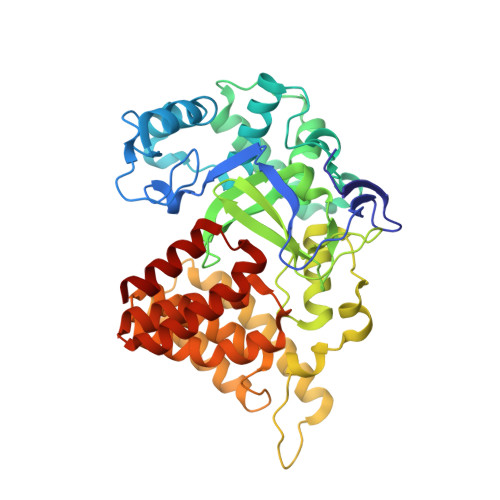
    Protein lysine methyltransferases (KMTs) have emerged as important regulators of epigenetic signaling. These enzymes catalyze the transfer of donor methyl groups from the cofactor S-adenosylmethionine to specific acceptor lysine residues on histones, leading to changes in chromatin structure and transcriptional regulation. These enzymes also methylate an array of nonhistone proteins, suggesting additional mechanisms by which they influence cellular physiology. SMYD2 is reported to be an oncogenic methyltransferase that represses the functional activity of the tumor suppressor proteins p53 and RB. HTS screening led to identification of five distinct substrate-competitive chemical series. Determination of liganded crystal structures of SMYD2 contributed significantly to "hit-to-lead" design efforts, culminating in the creation of potent and selective inhibitors that were used to understand the functional consequences of SMYD2 inhibition. Taken together, these results have broad implications for inhibitor design against KMTs and clearly demonstrate the potential for developing novel therapies against these enzymes.


  • Organizational Affiliation
    • Structure and Biophysics, Discovery Sciences, AstraZeneca, Mereside , Alderley Park, Cheshire, SK10 4TG United Kingdom.

Macromolecule Content 

  • Total Structure Weight: 50.85 kDa 
  • Atom Count: 3,888 
  • Modeled Residue Count: 429 
  • Deposited Residue Count: 429 
  • Unique protein chains: 1

Macromolecules

Find similar proteins by:|  3D Structure
Entity ID: 1
MoleculeChains  Sequence LengthOrganismDetailsImage
N-lysine methyltransferase SMYD2429Homo sapiensMutation(s): 1 
Gene Names: SMYD2KMT3C
EC: 2.1.1 (PDB Primary Data), 2.1.1.43 (PDB Primary Data), 2.1.1.354 (UniProt)
UniProt & NIH Common Fund Data Resources
Find proteins for Q9NRG4 (Homo sapiens)
Explore Q9NRG4 
Go to UniProtKB:  Q9NRG4
PHAROS:  Q9NRG4
GTEx:  ENSG00000143499 
Entity Groups
Sequence Clusters30% Identity50% Identity70% Identity90% Identity95% Identity100% Identity
UniProt GroupQ9NRG4
Sequence Annotations
Expand
Reference Sequence

Small Molecules

Ligands 4 Unique
IDChains Name / Formula / InChI Key2D Diagram3D Interactions
6T1

Query on 6T1


Download:Ideal Coordinates CCD File
G [auth A]2-[2-[1-[2-(3,4-dichlorophenyl)ethyl]azetidin-3-yl]oxyphenyl]-~{N}-(3-pyrrolidin-1-ylpropyl)pyridine-4-carboxamide
C30 H34 Cl2 N4 O2
AGSYPSPYUHOIIZ-UHFFFAOYSA-N
SAM

Query on SAM


Download:Ideal Coordinates CCD File
B [auth A]S-ADENOSYLMETHIONINE
C15 H22 N6 O5 S
MEFKEPWMEQBLKI-FCKMPRQPSA-N
BU3

Query on BU3


Download:Ideal Coordinates CCD File
C [auth A],
D [auth A],
E [auth A],
F [auth A]
(R,R)-2,3-BUTANEDIOL
C4 H10 O2
OWBTYPJTUOEWEK-QWWZWVQMSA-N
ZN

Query on ZN


Download:Ideal Coordinates CCD File
H [auth A],
I [auth A],
J [auth A]
ZINC ION
Zn
PTFCDOFLOPIGGS-UHFFFAOYSA-N

Experimental Data & Validation

Experimental Data

  • Method: X-RAY DIFFRACTION
  • Resolution: 1.93 Å
  • R-Value Free:  0.203 (Depositor), 0.201 (DCC) 
  • R-Value Work:  0.178 (Depositor) 
  • R-Value Observed: 0.179 (Depositor) 
Space Group: I 4
Unit Cell:
Length ( Å )Angle ( ˚ )
a = 155.865α = 90
b = 155.865β = 90
c = 52.477γ = 90
Software Package:
Software NamePurpose
BUSTERrefinement
XDSdata reduction
SCALAdata scaling
MOLREPphasing

Structure Validation

View Full Validation Report


Ligand Structure Quality Assessment 


Entry History 

Deposition Data

  • Released Date: 2016-12-07 
  • Deposition Author(s): Ferguson, A.

Revision History  (Full details and data files)

  • Version 1.0: 2016-12-07
    Type: Initial release
  • Version 1.1: 2017-01-04
    Changes: Database references
  • Version 1.2: 2023-09-27
    Changes: Data collection, Database references, Refinement description