5LP1 | pdb_00005lp1

CRYSTAL STRUCTURE OF HUMAN LIPOPROTEIN-ASSOCIATED PHOSPHOLIPASE A2 IN COMPLEX WITH A [1.1.1]BICYCLOPENTANE-CONTAINING INHIBITOR AT 1.91A RESOLUTION.


Experimental Data Snapshot

  • Method: X-RAY DIFFRACTION
  • Resolution: 1.91 Å
  • R-Value Free: 
    0.192 (Depositor), 0.203 (DCC) 
  • R-Value Work: 
    0.162 (Depositor), 0.173 (DCC) 
  • R-Value Observed: 
    0.164 (Depositor) 

Starting Model: other
View more details

wwPDB Validation 3D Report Full Report

Validation slider image for 5LP1

Ligand Structure Quality Assessment 


This is version 1.2 of the entry. See complete history

Literature

Investigation of a Bicyclo[1.1.1]pentane as a Phenyl Replacement within an LpPLA2 Inhibitor.

Measom, N.D.Down, K.D.Hirst, D.J.Jamieson, C.Manas, E.S.Patel, V.K.Somers, D.O.

(2017) ACS Med Chem Lett 8: 43-48

  • DOI: https://doi.org/10.1021/acsmedchemlett.6b00281
  • Primary Citation Related Structures: 
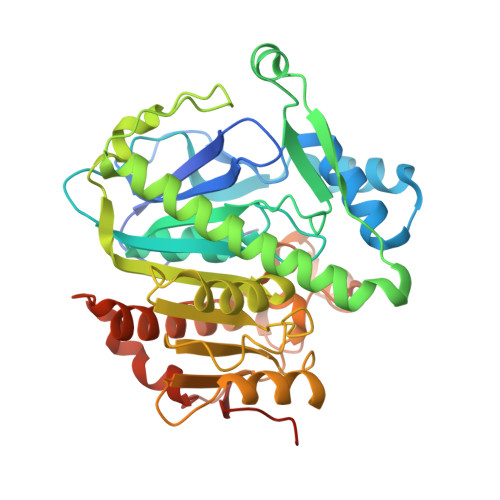
    5LP1

  • PubMed Abstract: 

    We describe the incorporation of a bicyclo[1.1.1]pentane moiety within two known LpPLA 2 inhibitors to act as bioisosteric phenyl replacements. An efficient synthesis to the target compounds was enabled with a dichlorocarbene insertion into a bicyclo[1.1.0]butane system being the key transformation. Potency, physicochemical, and X-ray crystallographic data were obtained to compare the known inhibitors to their bioisosteric counterparts, which showed the isostere was well tolerated and positively impacted on the physicochemical profile.


  • Organizational Affiliation
    • GlaxoSmithKline, Medicines Research Centre, Gunnels Wood Road, Stevenage, Hertfordshire, SG1 2NY, U.K.; Department of Pure and Applied Chemistry, University of Strathclyde, Thomas Graham Building, 295 Cathedral Street, Glasgow, G1 1XL, U.K.

Macromolecule Content 

  • Total Structure Weight: 44.93 kDa 
  • Atom Count: 3,420 
  • Modeled Residue Count: 370 
  • Deposited Residue Count: 388 
  • Unique protein chains: 1

Macromolecules

Find similar proteins by:|  3D Structure
Entity ID: 1
MoleculeChains  Sequence LengthOrganismDetailsImage
Platelet-activating factor acetylhydrolase388Homo sapiensMutation(s): 0 
Gene Names: PLA2G7PAFAH
EC: 3.1.1.47
UniProt & NIH Common Fund Data Resources
Find proteins for Q13093 (Homo sapiens)
Explore Q13093 
Go to UniProtKB:  Q13093
PHAROS:  Q13093
GTEx:  ENSG00000146070 
Entity Groups
Sequence Clusters30% Identity50% Identity70% Identity90% Identity95% Identity100% Identity
UniProt GroupQ13093
Sequence Annotations
Expand
Reference Sequence

Small Molecules

Ligands 2 Unique
IDChains Name / Formula / InChI Key2D Diagram3D Interactions
71H

Query on 71H


Download:Ideal Coordinates CCD File
D [auth A]~{N}-[2-(diethylamino)ethyl]-2-[2-[(4-fluorophenyl)methylsulfanyl]-4-oxidanylidene-5~{H}-cyclopenta[d]pyrimidin-1-yl]-~{N}-[[3-[4-(trifluoromethyl)phenyl]-1-bicyclo[1.1.1]pentanyl]methyl]ethanamide
C35 H38 F4 N4 O2 S
SHVFOCPBEIJQSO-AQOUDTPCSA-N
CL

Query on CL


Download:Ideal Coordinates CCD File
B [auth A],
C [auth A]
CHLORIDE ION
Cl
VEXZGXHMUGYJMC-UHFFFAOYSA-M

Experimental Data & Validation

Experimental Data

  • Method: X-RAY DIFFRACTION
  • Resolution: 1.91 Å
  • R-Value Free:  0.192 (Depositor), 0.203 (DCC) 
  • R-Value Work:  0.162 (Depositor), 0.173 (DCC) 
  • R-Value Observed: 0.164 (Depositor) 
Space Group: C 1 2 1
Unit Cell:
Length ( Å )Angle ( ˚ )
a = 100.28α = 90
b = 91.56β = 111.88
c = 51.65γ = 90
Software Package:
Software NamePurpose
XDSdata reduction
Aimlessdata scaling
REFMACphasing
REFMACrefinement
PDB_EXTRACTdata extraction

Structure Validation

View Full Validation Report


Ligand Structure Quality Assessment 


Entry History 

Deposition Data

  • Released Date: 2017-02-01 
  • Deposition Author(s): Somers, D.

Revision History  (Full details and data files)

  • Version 1.0: 2017-02-01
    Type: Initial release
  • Version 1.1: 2019-06-12
    Changes: Data collection, Structure summary
  • Version 1.2: 2024-05-01
    Changes: Data collection, Database references, Refinement description