5MR3

Crystal structure of red abalone egg VERL repeat 2 with linker in complex with sperm lysin at 1.8 A resolution


Experimental Data Snapshot

  • Method: X-RAY DIFFRACTION
  • Resolution: 1.80 Å
  • R-Value Free: 
    0.227 (Depositor), 0.230 (DCC) 
  • R-Value Work: 
    0.201 (Depositor), 0.200 (DCC) 
  • R-Value Observed: 
    0.202 (Depositor) 

Starting Models: experimental
View more details

wwPDB Validation   3D Report Full Report


Ligand Structure Quality Assessment 

Created with Raphaël 2.3.0Worse 01 BetterLigand structure goodness of fit to experimental dataBest fitted NAGClick on this verticalbar to view details

This is version 2.5 of the entry. See complete history


Literature

Structural Basis of Egg Coat-Sperm Recognition at Fertilization.

Raj, I.Sadat Al Hosseini, H.Dioguardi, E.Nishimura, K.Han, L.Villa, A.de Sanctis, D.Jovine, L.

(2017) Cell 169: 1315-1326.e17

  • DOI: https://doi.org/10.1016/j.cell.2017.05.033
  • Primary Citation of Related Structures:  
    5II4, 5II5, 5II6, 5II7, 5II8, 5II9, 5IIA, 5IIB, 5IIC, 5MR2, 5MR3

  • PubMed Abstract: 

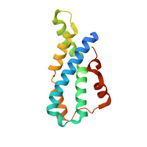
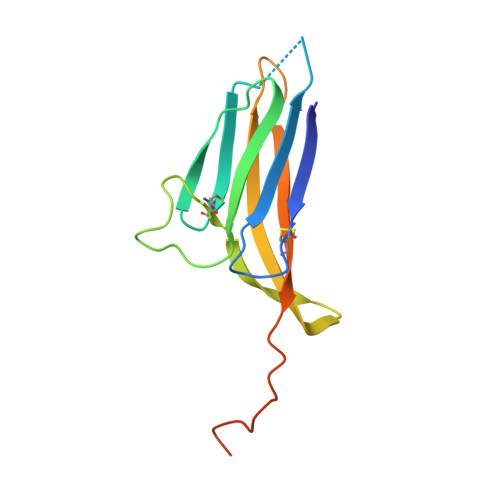
    Recognition between sperm and the egg surface marks the beginning of life in all sexually reproducing organisms. This fundamental biological event depends on the species-specific interaction between rapidly evolving counterpart molecules on the gametes. We report biochemical, crystallographic, and mutational studies of domain repeats 1-3 of invertebrate egg coat protein VERL and their interaction with cognate sperm protein lysin. VERL repeats fold like the functionally essential N-terminal repeat of mammalian sperm receptor ZP2, whose structure is also described here. Whereas sequence-divergent repeat 1 does not bind lysin, repeat 3 binds it non-species specifically via a high-affinity, largely hydrophobic interface. Due to its intermediate binding affinity, repeat 2 selectively interacts with lysin from the same species. Exposure of a highly positively charged surface of VERL-bound lysin suggests that complex formation both disrupts the organization of egg coat filaments and triggers their electrostatic repulsion, thereby opening a hole for sperm penetration and fusion.


  • Organizational Affiliation

    Department of Biosciences and Nutrition and Center for Innovative Medicine, Karolinska Institutet, Huddinge, SE-141 83, Sweden.


Macromolecules
Find similar proteins by:  (by identity cutoff)  |  3D Structure
Entity ID: 1
MoleculeChains Sequence LengthOrganismDetailsImage
Egg-lysin
A, C, E, G
136Haliotis rufescensMutation(s): 0 
UniProt
Find proteins for P04552 (Haliotis rufescens)
Explore P04552 
Go to UniProtKB:  P04552
Entity Groups  
Sequence Clusters30% Identity50% Identity70% Identity90% Identity95% Identity100% Identity
UniProt GroupP04552
Sequence Annotations
Expand
  • Reference Sequence
Find similar proteins by:  (by identity cutoff)  |  3D Structure
Entity ID: 2
MoleculeChains Sequence LengthOrganismDetailsImage
Vitelline envelope sperm lysin receptor
B, D, F, H
134Haliotis rufescensMutation(s): 3 
Gene Names: VERL
UniProt
Find proteins for Q8WR62 (Haliotis rufescens)
Explore Q8WR62 
Go to UniProtKB:  Q8WR62
Entity Groups  
Sequence Clusters30% Identity50% Identity70% Identity90% Identity95% Identity100% Identity
UniProt GroupQ8WR62
Glycosylation
Glycosylation Sites: 1
Sequence Annotations
Expand
  • Reference Sequence
Small Molecules
Ligands 4 Unique
IDChains Name / Formula / InChI Key2D Diagram3D Interactions
NAG
Query on NAG

Download Ideal Coordinates CCD File 
BA [auth H],
N [auth B],
S [auth D],
W [auth F]
2-acetamido-2-deoxy-beta-D-glucopyranose
C8 H15 N O6
OVRNDRQMDRJTHS-FMDGEEDCSA-N
PGE
Query on PGE

Download Ideal Coordinates CCD File 
X [auth G],
Y [auth G]
TRIETHYLENE GLYCOL
C6 H14 O4
ZIBGPFATKBEMQZ-UHFFFAOYSA-N
GOL
Query on GOL

Download Ideal Coordinates CCD File 
AA [auth H]
I [auth A]
J [auth B]
K [auth B]
M [auth B]
GLYCEROL
C3 H8 O3
PEDCQBHIVMGVHV-UHFFFAOYSA-N
CL
Query on CL

Download Ideal Coordinates CCD File 
L [auth B],
V [auth F]
CHLORIDE ION
Cl
VEXZGXHMUGYJMC-UHFFFAOYSA-M
Experimental Data & Validation

Experimental Data

  • Method: X-RAY DIFFRACTION
  • Resolution: 1.80 Å
  • R-Value Free:  0.227 (Depositor), 0.230 (DCC) 
  • R-Value Work:  0.201 (Depositor), 0.200 (DCC) 
  • R-Value Observed: 0.202 (Depositor) 
Space Group: P 1 21 1
Unit Cell:
Length ( Å )Angle ( ˚ )
a = 75.87α = 90
b = 87.86β = 100.93
c = 92.55γ = 90
Software Package:
Software NamePurpose
PHENIXrefinement
XDSdata reduction
XDSdata scaling
PHASERphasing
Cootmodel building

Structure Validation

View Full Validation Report


Ligand Structure Quality Assessment 

Created with Raphaël 2.3.0Worse 01 BetterLigand structure goodness of fit to experimental dataBest fitted NAGClick on this verticalbar to view details

Entry History & Funding Information

Deposition Data


Funding OrganizationLocationGrant Number
Karolinska InstitutetSweden--
Swedish Research CouncilSweden2012-5093
Goran Gustafsson Foundation for Research in Natural Sciences and MedicineSweden--
Sven and Ebba-Christina Hagberg foundationSweden--
European Molecular Biology Organization--
European UnionERC 260759

Revision History  (Full details and data files)

  • Version 1.0: 2017-06-14
    Type: Initial release
  • Version 1.1: 2017-06-28
    Changes: Database references
  • Version 2.0: 2017-07-26
    Changes: Advisory, Database references, Polymer sequence, Source and taxonomy, Structure summary
  • Version 2.1: 2017-09-06
    Changes: Author supporting evidence
  • Version 2.2: 2017-11-22
    Changes: Database references
  • Version 2.3: 2020-07-29
    Type: Remediation
    Reason: Carbohydrate remediation
    Changes: Data collection, Derived calculations, Structure summary
  • Version 2.4: 2024-01-17
    Changes: Data collection, Database references, Refinement description, Structure summary
  • Version 2.5: 2024-10-16
    Changes: Structure summary