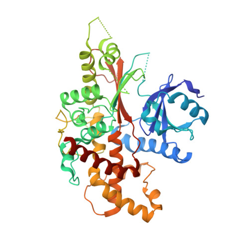
5MW8

INOSITOL 1,3,4,5,6-PENTAKISPHOSPHATE 2-KINASE FROM M. MUSCULUS IN COMPLEX WITH ATP and IP5


Experimental Data Snapshot

  • Method: X-RAY DIFFRACTION
  • Resolution: 2.40 Å
  • R-Value Free: 0.271 
  • R-Value Work: 0.241 
  • R-Value Observed: 0.243 

wwPDB Validation   3D Report Full Report


Ligand Structure Quality Assessment 


This is version 1.2 of the entry. See complete history


Literature

The crystal structure of mammalian inositol 1,3,4,5,6-pentakisphosphate 2-kinase reveals a new zinc-binding site and key features for protein function.

Franco-Echevarria, E.Sanz-Aparicio, J.Brearley, C.A.Gonzalez-Rubio, J.M.Gonzalez, B.

(2017) J Biol Chem 292: 10534-10548

  • DOI: https://doi.org/10.1074/jbc.M117.780395
  • Primary Citation of Related Structures:  
    5MW8, 5MWL, 5MWM

  • PubMed Abstract: 

    Inositol 1,3,4,5,6-pentakisphosphate 2-kinases (IP 5 2-Ks) are part of a family of enzymes in charge of synthesizing inositol hexakisphosphate (IP 6 ) in eukaryotic cells. This protein and its product IP 6 present many roles in cells, participating in mRNA export, embryonic development, and apoptosis. We reported previously that the full-length IP 5 2-K from Arabidopsis thaliana is a zinc metallo-enzyme, including two separated lobes (the N- and C-lobes). We have also shown conformational changes in IP 5 2-K and have identified the residues involved in substrate recognition and catalysis. However, the specific features of mammalian IP 5 2-Ks remain unknown. To this end, we report here the first structure for a murine IP 5 2-K in complex with ATP/IP 5 or IP 6 Our structural findings indicated that the general folding in N- and C-lobes is conserved with A. thaliana IP 5 2-K. A helical scaffold in the C-lobe constitutes the inositol phosphate-binding site, which, along with the participation of the N-lobe, endows high specificity to this protein. However, we also noted large structural differences between the orthologues from these two eukaryotic kingdoms. These differences include a novel zinc-binding site and regions unique to the mammalian IP 5 2-K, as an unexpected basic patch on the protein surface. In conclusion, our findings have uncovered distinct features of a mammalian IP 5 2-K and set the stage for investigations into protein-protein or protein-RNA interactions important for IP 5 2-K function and activity.


  • Organizational Affiliation

    From the Departamento de Cristalografía y Biología Estructural, Instituto de Química-Física "Rocasolano," Consejo Superior de Investigaciones Científicas, Serrano 119, 28006 Madrid, Spain and.


Macromolecules
Find similar proteins by:  (by identity cutoff)  |  3D Structure
Entity ID: 1
MoleculeChains Sequence LengthOrganismDetailsImage
Inositol-pentakisphosphate 2-kinase471Mus musculusMutation(s): 0 
Gene Names: Ippk
EC: 2.7.1.158
UniProt & NIH Common Fund Data Resources
Find proteins for Q6P1C1 (Mus musculus)
Explore Q6P1C1 
Go to UniProtKB:  Q6P1C1
IMPC:  MGI:1922928
Entity Groups  
Sequence Clusters30% Identity50% Identity70% Identity90% Identity95% Identity100% Identity
UniProt GroupQ6P1C1
Sequence Annotations
Expand
  • Reference Sequence
Small Molecules
Ligands 5 Unique
IDChains Name / Formula / InChI Key2D Diagram3D Interactions
5MY
Query on 5MY

Download Ideal Coordinates CCD File 
C [auth A]MYO-INOSITOL-(1,3,4,5,6)-PENTAKISPHOSPHATE
C6 H17 O21 P5
CTPQAXVNYGZUAJ-KXXVROSKSA-N
ATP
Query on ATP

Download Ideal Coordinates CCD File 
B [auth A]ADENOSINE-5'-TRIPHOSPHATE
C10 H16 N5 O13 P3
ZKHQWZAMYRWXGA-KQYNXXCUSA-N
ZN
Query on ZN

Download Ideal Coordinates CCD File 
D [auth A]ZINC ION
Zn
PTFCDOFLOPIGGS-UHFFFAOYSA-N
ACT
Query on ACT

Download Ideal Coordinates CCD File 
G [auth A]ACETATE ION
C2 H3 O2
QTBSBXVTEAMEQO-UHFFFAOYSA-M
MG
Query on MG

Download Ideal Coordinates CCD File 
E [auth A],
F [auth A]
MAGNESIUM ION
Mg
JLVVSXFLKOJNIY-UHFFFAOYSA-N
Experimental Data & Validation

Experimental Data

  • Method: X-RAY DIFFRACTION
  • Resolution: 2.40 Å
  • R-Value Free: 0.271 
  • R-Value Work: 0.241 
  • R-Value Observed: 0.243 
  • Space Group: P 1 21 1
Unit Cell:
Length ( Å )Angle ( ˚ )
a = 60.162α = 90
b = 71.495β = 111.37
c = 61.197γ = 90
Software Package:
Software NamePurpose
REFMACrefinement
XDSdata reduction
XDSdata scaling
Aimlessdata scaling
Cootmodel building
PHENIXphasing
RESOLVEphasing

Structure Validation

View Full Validation Report


Ligand Structure Quality Assessment 


Entry History & Funding Information

Deposition Data


Funding OrganizationLocationGrant Number
CSICSpainBFU2014-53762-P

Revision History  (Full details and data files)

  • Version 1.0: 2017-05-10
    Type: Initial release
  • Version 1.1: 2017-07-05
    Changes: Database references
  • Version 1.2: 2024-05-08
    Changes: Data collection, Database references, Derived calculations