5NHC | pdb_00005nhc

Crystal structure of xylose isomerase from Piromyces E2 in complex with two Co2+ ions and xylulose


Experimental Data Snapshot

  • Method: X-RAY DIFFRACTION
  • Resolution: 1.93 Å
  • R-Value Free: 
    0.188 (Depositor), 0.167 (DCC) 
  • R-Value Work: 
    0.157 (Depositor) 
  • R-Value Observed: 
    0.158 (Depositor) 

Starting Model: experimental
View more details

wwPDB Validation 3D Report Full Report

Validation slider image for 5NHC

Ligand Structure Quality Assessment 


This is version 1.5 of the entry. See complete history

Literature

Metal Dependence of the Xylose Isomerase from Piromyces sp. E2 Explored by Activity Profiling and Protein Crystallography.

Lee, M.Rozeboom, H.J.de Waal, P.P.de Jong, R.M.Dudek, H.M.Janssen, D.B.

(2017) Biochemistry 56: 5991-6005

  • DOI: https://doi.org/10.1021/acs.biochem.7b00777
  • Primary Citation Related Structures: 
    5NH4, 5NH5, 5NH6, 5NH7, 5NH8, 5NH9, 5NHA, 5NHB, 5NHC, 5NHD, 5NHE, 5NHM

  • PubMed Abstract: 

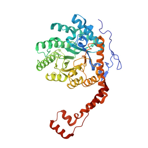
    Xylose isomerase from Piromyces sp. E2 (PirXI) can be used to equip Saccharomyces cerevisiae with the capacity to ferment xylose to ethanol. The biochemical properties and structure of the enzyme have not been described even though its metal content, catalytic parameters, and expression level are critical for rapid xylose utilization. We have isolated the enzyme after high-level expression in Escherichia coli, analyzed the metal dependence of its catalytic properties, and determined 12 crystal structures in the presence of different metals, substrates, and substrate analogues. The activity assays revealed that various bivalent metals can activate PirXI for xylose isomerization. Among these metals, Mn 2+ is the most favorable for catalytic activity. Furthermore, the enzyme shows the highest affinity for Mn 2+ , which was established by measuring the activation constants (K act ) for different metals. Metal analysis of the purified enzyme showed that in vivo the enzyme binds a mixture of metals that is determined by metal availability as well as affinity, indicating that the native metal composition can influence activity. The crystal structures show the presence of an active site similar to that of other xylose isomerases, with a d-xylose binding site containing two tryptophans and a catalytic histidine, as well as two metal binding sites that are formed by carboxylate groups of conserved aspartates and glutamates. The binding positions and conformations of the metal-coordinating residues varied slightly for different metals, which is hypothesized to contribute to the observed metal dependence of the isomerase activity.


  • Organizational Affiliation
    • Biochemical Laboratory, Groningen Biomolecular Sciences and Biotechnology Institute, University of Groningen , Nijenborgh 4, 9747 AG Groningen, The Netherlands.

Macromolecule Content 

  • Total Structure Weight: 199.43 kDa 
  • Atom Count: 16,258 
  • Modeled Residue Count: 1,742 
  • Deposited Residue Count: 1,748 
  • Unique protein chains: 1

Macromolecules

Find similar proteins by:|  3D Structure
Entity ID: 1
MoleculeChains  Sequence LengthOrganismDetailsImage
Xylose isomerase
A, B, C, D
437Piromyces sp. E2Mutation(s): 0 
Gene Names: xylA
EC: 5.3.1.5
UniProt
Find proteins for A0ACD6B9F5 (Piromyces sp. (strain E2))
Explore A0ACD6B9F5 
Go to UniProtKB:  A0ACD6B9F5
Entity Groups
Sequence Clusters30% Identity50% Identity70% Identity90% Identity95% Identity100% Identity
UniProt GroupA0ACD6B9F5
Sequence Annotations
Expand
Reference Sequence

Small Molecules

Ligands 3 Unique
IDChains Name / Formula / InChI Key2D Diagram3D Interactions
XUL

Query on XUL


Download:Ideal Coordinates CCD File
G [auth A],
K [auth B],
O [auth C],
S [auth D]
D-XYLULOSE
C5 H10 O5
ZAQJHHRNXZUBTE-WUJLRWPWSA-N
HYP

Query on HYP


Download:Ideal Coordinates CCD File
H [auth A],
L [auth B],
P [auth C],
T [auth D]
4-HYDROXYPROLINE
C5 H9 N O3
PMMYEEVYMWASQN-DMTCNVIQSA-N
CO

Query on CO


Download:Ideal Coordinates CCD File
E [auth A]
F [auth A]
I [auth B]
J [auth B]
M [auth C]
E [auth A],
F [auth A],
I [auth B],
J [auth B],
M [auth C],
N [auth C],
Q [auth D],
R [auth D]
COBALT (II) ION
Co
XLJKHNWPARRRJB-UHFFFAOYSA-N

Experimental Data & Validation

Experimental Data

  • Method: X-RAY DIFFRACTION
  • Resolution: 1.93 Å
  • R-Value Free:  0.188 (Depositor), 0.167 (DCC) 
  • R-Value Work:  0.157 (Depositor) 
  • R-Value Observed: 0.158 (Depositor) 
Space Group: P 1
Unit Cell:
Length ( Å )Angle ( ˚ )
a = 79.249α = 115.72
b = 79.332β = 89.39
c = 91.359γ = 116.87
Software Package:
Software NamePurpose
REFMACrefinement
XDSdata reduction
Aimlessdata scaling
PHASERphasing

Structure Validation

View Full Validation Report


Ligand Structure Quality Assessment 


Entry History 

Deposition Data

Revision History  (Full details and data files)

  • Version 1.0: 2017-11-01
    Type: Initial release
  • Version 1.1: 2017-11-15
    Changes: Database references
  • Version 1.2: 2017-11-22
    Changes: Database references
  • Version 1.3: 2018-08-29
    Changes: Data collection, Database references
  • Version 1.4: 2019-11-20
    Changes: Derived calculations
  • Version 1.5: 2024-05-01
    Changes: Data collection, Database references, Derived calculations, Refinement description