5ODW

Structure of the FpvAI-pyocin S2 complex


Experimental Data Snapshot

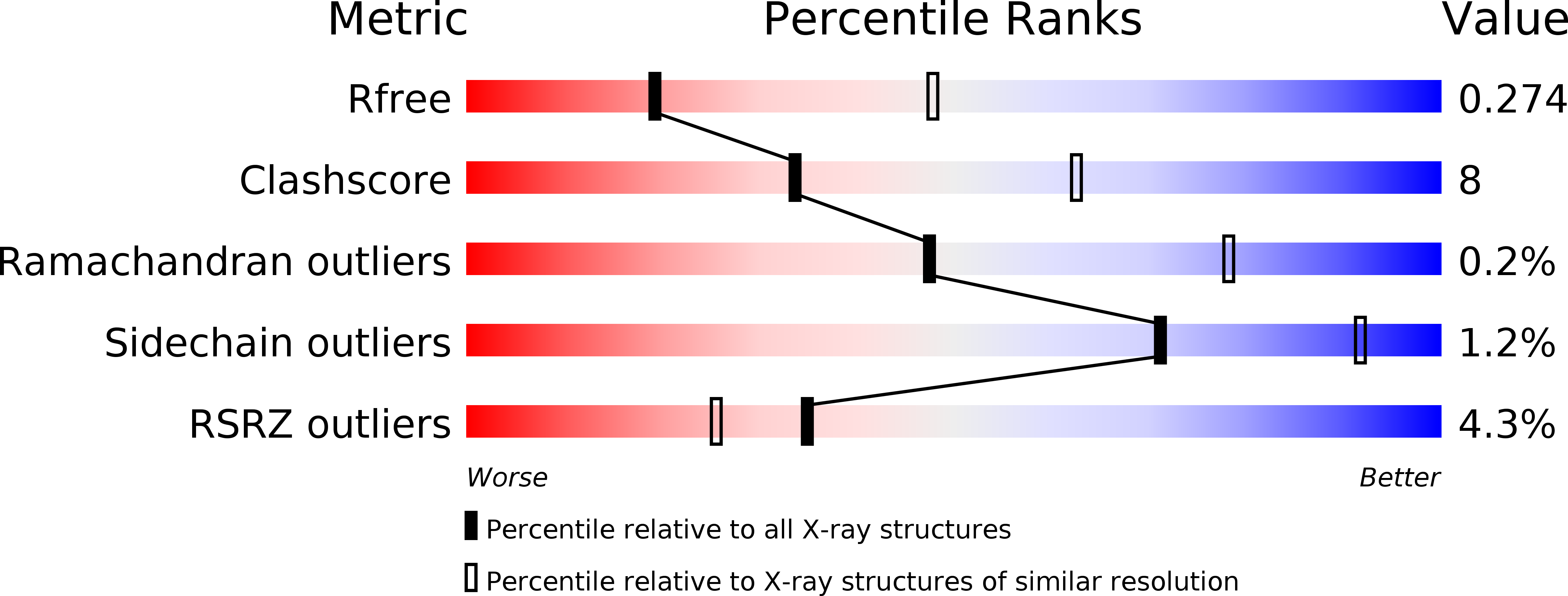
  • Method: X-RAY DIFFRACTION
  • Resolution: 2.80 Å
  • R-Value Free: 0.262 
  • R-Value Work: 0.212 
  • R-Value Observed: 0.214 

Starting Model: experimental
View more details

wwPDB Validation   3D Report Full Report


This is version 1.2 of the entry. See complete history


Literature

Exploitation of an iron transporter for bacterial protein antibiotic import.

White, P.Joshi, A.Rassam, P.Housden, N.G.Kaminska, R.Goult, J.D.Redfield, C.McCaughey, L.C.Walker, D.Mohammed, S.Kleanthous, C.

(2017) Proc Natl Acad Sci U S A 114: 12051-12056

  • DOI: https://doi.org/10.1073/pnas.1713741114
  • Primary Citation of Related Structures:  
    5ODW

  • PubMed Abstract: 

    Unlike their descendants, mitochondria and plastids, bacteria do not have dedicated protein import systems. However, paradoxically, import of protein bacteriocins, the mechanisms of which are poorly understood, underpins competition among pathogenic and commensal bacteria alike. Here, using X-ray crystallography, isothermal titration calorimetry, confocal fluorescence microscopy, and in vivo photoactivatable cross-linking of stalled translocation intermediates, we demonstrate how the iron transporter FpvAI in the opportunistic pathogen Pseudomonas aeruginosa is hijacked to translocate the bacteriocin pyocin S2 (pyoS2) across the outer membrane (OM). FpvAI is a TonB-dependent transporter (TBDT) that actively imports the small siderophore ferripyoverdine (Fe-Pvd) by coupling to the proton motive force (PMF) via the inner membrane (IM) protein TonB1. The crystal structure of the N-terminal domain of pyoS2 (pyoS2 NTD ) bound to FpvAI ( K d = 240 pM) reveals that the pyocin mimics Fe-Pvd, inducing the same conformational changes in the receptor. Mimicry leads to fluorescently labeled pyoS2 NTD being imported into FpvAI-expressing P. aeruginosa cells by a process analogous to that used by bona fide TBDT ligands. PyoS2 NTD induces unfolding by TonB1 of a force-labile portion of the plug domain that normally occludes the central channel of FpvAI. The pyocin is then dragged through this narrow channel following delivery of its own TonB1-binding epitope to the periplasm. Hence, energized nutrient transporters in bacteria also serve as rudimentary protein import systems, which, in the case of FpvAI, results in a protein antibiotic 60-fold bigger than the transporter's natural substrate being translocated across the OM.


  • Organizational Affiliation

    Department of Biochemistry, University of Oxford, Oxford OX1 3QU, United Kingdom.


Macromolecules
Find similar proteins by:  (by identity cutoff)  |  3D Structure
Entity ID: 1
MoleculeChains Sequence LengthOrganismDetailsImage
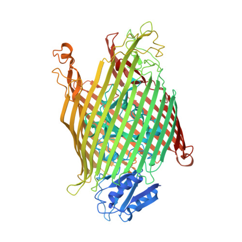
Ferripyoverdine receptor
A, B
815Pseudomonas aeruginosa PAO1Mutation(s): 0 
Gene Names: fpvAPA2398
Membrane Entity: Yes 
UniProt
Find proteins for P48632 (Pseudomonas aeruginosa (strain ATCC 15692 / DSM 22644 / CIP 104116 / JCM 14847 / LMG 12228 / 1C / PRS 101 / PAO1))
Explore P48632 
Go to UniProtKB:  P48632
Entity Groups  
Sequence Clusters30% Identity50% Identity70% Identity90% Identity95% Identity100% Identity
UniProt GroupP48632
Sequence Annotations
Expand
  • Reference Sequence
Find similar proteins by:  (by identity cutoff)  |  3D Structure
Entity ID: 2
MoleculeChains Sequence LengthOrganismDetailsImage
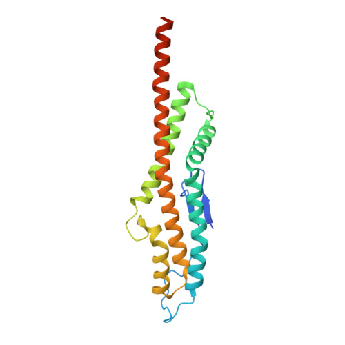
Pyocin-S2
C, D
217Pseudomonas aeruginosa PAO1Mutation(s): 0 
Gene Names: pys2PA1150
EC: 3.1
UniProt
Find proteins for Q06584 (Pseudomonas aeruginosa (strain ATCC 15692 / DSM 22644 / CIP 104116 / JCM 14847 / LMG 12228 / 1C / PRS 101 / PAO1))
Explore Q06584 
Go to UniProtKB:  Q06584
Entity Groups  
Sequence Clusters30% Identity50% Identity70% Identity90% Identity95% Identity100% Identity
UniProt GroupQ06584
Sequence Annotations
Expand
  • Reference Sequence
Small Molecules
Ligands 1 Unique
IDChains Name / Formula / InChI Key2D Diagram3D Interactions
SO4
Query on SO4

Download Ideal Coordinates CCD File 
E [auth A]
F [auth A]
G [auth A]
H [auth A]
I [auth A]
E [auth A],
F [auth A],
G [auth A],
H [auth A],
I [auth A],
J [auth B],
K [auth B],
L [auth B],
M [auth B],
N [auth B]
SULFATE ION
O4 S
QAOWNCQODCNURD-UHFFFAOYSA-L
Experimental Data & Validation

Experimental Data

  • Method: X-RAY DIFFRACTION
  • Resolution: 2.80 Å
  • R-Value Free: 0.262 
  • R-Value Work: 0.212 
  • R-Value Observed: 0.214 
  • Space Group: I 2 2 2
Unit Cell:
Length ( Å )Angle ( ˚ )
a = 115.524α = 90
b = 209.39β = 90
c = 215.845γ = 90
Software Package:
Software NamePurpose
BUSTERrefinement
XDSdata reduction
Aimlessdata scaling
PHASERphasing

Structure Validation

View Full Validation Report


Entry History & Funding Information

Deposition Data


Funding OrganizationLocationGrant Number
Wellcome TrustUnited Kingdom201505/Z/16/Z

Revision History  (Full details and data files)

  • Version 1.0: 2017-11-08
    Type: Initial release
  • Version 1.1: 2017-11-15
    Changes: Database references
  • Version 1.2: 2024-01-17
    Changes: Data collection, Database references, Refinement description