5SZG | pdb_00005szg

Structure of the bMERB domain of Mical-3


Experimental Data Snapshot

  • Method: X-RAY DIFFRACTION
  • Resolution: 2.70 Å
  • R-Value Free: 
    0.282 (Depositor), 0.291 (DCC) 
  • R-Value Work: 
    0.251 (Depositor), 0.263 (DCC) 
  • R-Value Observed: 
    0.252 (Depositor) 

wwPDB Validation 3D Report Full Report

Validation slider image for 5SZG

This is version 1.4 of the entry. See complete history

Literature

bMERB domains are bivalent Rab8 family effectors evolved by gene duplication.

Rai, A.Oprisko, A.Campos, J.Fu, Y.Friese, T.Itzen, A.Goody, R.S.Gazdag, E.M.Muller, M.P.

(2016) Elife 5

  • DOI: https://doi.org/10.7554/eLife.18675
  • Primary Citation Related Structures: 
    5LPN, 5SZG, 5SZH, 5SZI, 5SZJ, 5SZK

  • PubMed Abstract: 

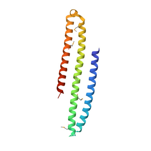
    In their active GTP-bound form, Rab proteins interact with proteins termed effector molecules. In this study, we have thoroughly characterized a Rab effector domain that is present in proteins of the Mical and EHBP families, both known to act in endosomal trafficking. Within our study, we show that these effectors display a preference for Rab8 family proteins (Rab8, 10, 13 and 15) and that some of the effector domains can bind two Rab proteins via separate binding sites. Structural analysis allowed us to explain the specificity towards Rab8 family members and the presence of two similar Rab binding sites that must have evolved via gene duplication. This study is the first to thoroughly characterize a Rab effector protein that contains two separate Rab binding sites within a single domain, allowing Micals and EHBPs to bind two Rabs simultaneously, thus suggesting previously unknown functions of these effector molecules in endosomal trafficking.


  • Organizational Affiliation
    • Department of Structural Biochemistry, Max Planck Institute of Molecular Physiology, Dortmund, Germany.

Macromolecule Content 

  • Total Structure Weight: 37.29 kDa 
  • Atom Count: 2,109 
  • Modeled Residue Count: 262 
  • Deposited Residue Count: 302 
  • Unique protein chains: 1

Macromolecules

Find similar proteins by:|  3D Structure
Entity ID: 1
MoleculeChains  Sequence LengthOrganismDetailsImage
Protein-methionine sulfoxide oxidase MICAL3
A, B
151Homo sapiensMutation(s): 0 
Gene Names: MICAL3KIAA0819KIAA1364
EC: 1.14.13 (PDB Primary Data), 1.14.13.225 (UniProt)
UniProt & NIH Common Fund Data Resources
Find proteins for Q7RTP6 (Homo sapiens)
Explore Q7RTP6 
Go to UniProtKB:  Q7RTP6
PHAROS:  Q7RTP6
GTEx:  ENSG00000243156 
Entity Groups
Sequence Clusters30% Identity50% Identity70% Identity90% Identity95% Identity100% Identity
UniProt GroupQ7RTP6
Sequence Annotations
Expand
Reference Sequence

Small Molecules

Modified Residues  1 Unique
IDChains TypeFormula2D DiagramParent
MSE
Query on MSE
A, B
L-PEPTIDE LINKINGC5 H11 N O2 SeMET

Experimental Data & Validation

Experimental Data

  • Method: X-RAY DIFFRACTION
  • Resolution: 2.70 Å
  • R-Value Free:  0.282 (Depositor), 0.291 (DCC) 
  • R-Value Work:  0.251 (Depositor), 0.263 (DCC) 
  • R-Value Observed: 0.252 (Depositor) 
Space Group: P 21 21 21
Unit Cell:
Length ( Å )Angle ( ˚ )
a = 51.858α = 90
b = 78.848β = 90
c = 95.557γ = 90
Software Package:
Software NamePurpose
REFMACrefinement
XDSdata reduction
XSCALEdata scaling
PHASERphasing

Structure Validation

View Full Validation Report


Entry History 

& Funding Information

Deposition Data


Funding OrganizationLocationGrant Number
German Research FoundationGermanySFB 642, grant A4
Max Planck SocietyGermany--

Revision History  (Full details and data files)

  • Version 1.0: 2016-08-24
    Type: Initial release
  • Version 1.1: 2016-09-07
    Changes: Database references
  • Version 1.2: 2016-09-14
    Changes: Database references
  • Version 1.3: 2017-09-06
    Changes: Author supporting evidence
  • Version 1.4: 2024-11-13
    Changes: Data collection, Database references, Refinement description, Structure summary