Download Mendeley
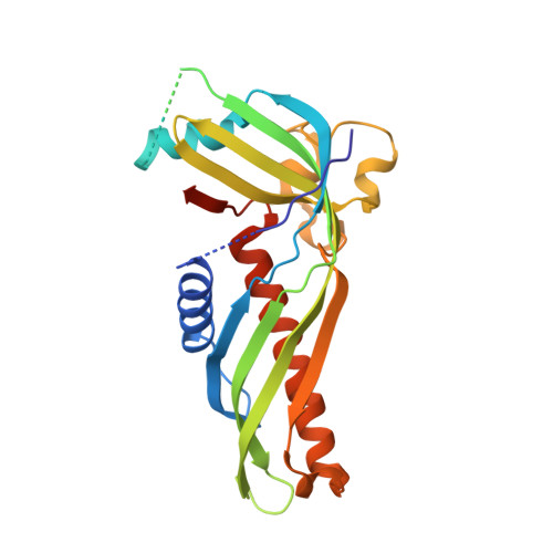
Crystal structure of Mdm12 and combinatorial reconstitution of Mdm12/Mmm1 ERMES complexes for structural studies.
AhYoung, A.P., Lu, B., Cascio, D., Egea, P.F.(2017) Biochem Biophys Res Commun 488: 129-135
- PubMed: 28479252 Search on PubMedSearch on PubMed Central
- DOI: https://doi.org/10.1016/j.bbrc.2017.05.021
- Primary Citation Related Structures:
5VKZ - PubMed Abstract:
Membrane contact sites between organelles serve as molecular hubs for the exchange of metabolites and signals. In yeast, the Endoplasmic Reticulum - Mitochondrion Encounter Structure (ERMES) tethers these two organelles likely to facilitate the non-vesicular exchange of essential phospholipids. Present in Fungi and Amoebas but not in Metazoans, ERMES is composed of five distinct subunits; among those, Mdm12, Mmm1 and Mdm34 each contain an SMP domain functioning as a lipid transfer module. We previously showed that the SMP domains of Mdm12 and Mmm1 form a hetero-tetramer. Here we describe our strategy to diversify the number of Mdm12/Mmm1 complexes suited for structural studies. We use sequence analysis of orthologues combined to protein engineering of disordered regions to guide the design of protein constructs and expand the repertoire of Mdm12/Mmm1 complexes more likely to crystallize. Using this combinatorial approach we report crystals of Mdm12/Mmm1 ERMES complexes currently diffracting to 4.5 Å resolution and a new structure of Mdm12 solved at 4.1 Å resolution. Our structure reveals a monomeric form of Mdm12 with a conformationally dynamic N-terminal β-strand; it differs from a previously reported homodimeric structure where the N-terminal β strands where swapped to promote dimerization. Based on our electron microscopy data, we propose a refined pseudo-atomic model of the Mdm12/Mmm1 complex that agrees with our crystallographic and small-angle X-ray scattering (SAXS) solution data.
- Department of Biological Chemistry, David Geffen School of Medicine, UCLA, Los Angeles, USA.
Organizational Affiliation: