5VT4 | pdb_00005vt4

Sco GlgEI-V279S in complex with a pyrolidene-based methyl-phosphonate compound


Experimental Data Snapshot

  • Method: X-RAY DIFFRACTION
  • Resolution: 3.21 Å
  • R-Value Free: 
    0.239 (Depositor), 0.243 (DCC) 
  • R-Value Work: 
    0.207 (Depositor), 0.215 (DCC) 
  • R-Value Observed: 
    0.208 (Depositor) 

Starting Model: experimental
View more details

wwPDB Validation 3D Report Full Report

Validation slider image for 5VT4

Ligand Structure Quality Assessment 


This is version 2.1 of the entry. See complete history

Literature

Zwitterionic pyrrolidene-phosphonates: inhibitors of the glycoside hydrolase-like phosphorylase Streptomyces coelicolor GlgEI-V279S.

Veleti, S.K.Petit, C.Ronning, D.R.Sucheck, S.J.

(2017) Org Biomol Chem 15: 3884-3891

  • DOI: https://doi.org/10.1039/c7ob00388a
  • Primary Citation Related Structures: 
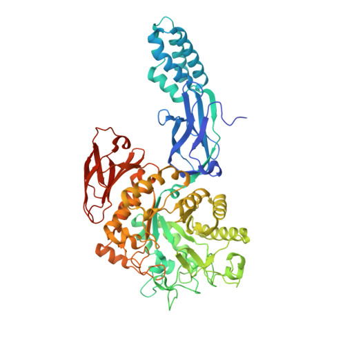
    5VSJ, 5VT4

  • PubMed Abstract: 

    We synthesized and evaluated new zwitterionic inhibitors against glycoside hydrolase-like phosphorylase Streptomyces coelicolor (Sco) GlgEI-V279S which plays a role in α-glucan biosynthesis. Sco GlgEI-V279S serves as a model enzyme for validated anti-tuberculosis (TB) target Mycobacterium tuberculosis (Mtb) GlgE. Pyrrolidine inhibitors 5 and 6 were designed based on transition state considerations and incorporate a phosphonate on the pyrrolidine moiety to expand the interaction network between the inhibitor and the enzyme active site. Compounds 5 and 6 inhibited Sco GlgEI-V279S with K i = 45 ± 4 μM and 95 ± 16 μM, respectively, and crystal structures of Sco GlgE-V279S-5 and Sco GlgE-V279S-6 were obtained at a 3.2 Å and 2.5 Å resolution, respectively.


  • Organizational Affiliation
    • Department of Chemistry and Biochemistry and School of Green Chemistry and Engineering, The University of Toledo, 2801 W. Bancroft Street, Toledo, Ohio 43606, USA. steve.sucheck@utoledo.edu donald.ronning@utoledo.edu.

Macromolecule Content 

  • Total Structure Weight: 298.39 kDa 
  • Atom Count: 20,458 
  • Modeled Residue Count: 2,580 
  • Deposited Residue Count: 2,652 
  • Unique protein chains: 1

Macromolecules

Find similar proteins by:|  3D Structure
Entity ID: 1
MoleculeChains  Sequence LengthOrganismDetailsImage
Alpha-1,4-glucan:maltose-1-phosphate maltosyltransferase 1
A, B, C, D
663Streptomyces coelicolor A3(2)Mutation(s): 1 
Gene Names: glgE1pep1pep1Apep1ISCO5443SC6A11.19c
EC: 2.4.99.16
UniProt
Find proteins for Q9L1K2 (Streptomyces coelicolor (strain ATCC BAA-471 / A3(2) / M145))
Explore Q9L1K2 
Go to UniProtKB:  Q9L1K2
Entity Groups
Sequence Clusters30% Identity50% Identity70% Identity90% Identity95% Identity100% Identity
UniProt GroupQ9L1K2
Sequence Annotations
Expand
Reference Sequence

Experimental Data & Validation

Experimental Data

  • Method: X-RAY DIFFRACTION
  • Resolution: 3.21 Å
  • R-Value Free:  0.239 (Depositor), 0.243 (DCC) 
  • R-Value Work:  0.207 (Depositor), 0.215 (DCC) 
  • R-Value Observed: 0.208 (Depositor) 
Space Group: P 41
Unit Cell:
Length ( Å )Angle ( ˚ )
a = 113.851α = 90
b = 113.851β = 90
c = 312.49γ = 90
Software Package:
Software NamePurpose
PHENIXrefinement
PHENIXmodel building
HKL-2000data scaling
HKL-2000data reduction

Structure Validation

View Full Validation Report


Ligand Structure Quality Assessment 


Entry History 

& Funding Information

Deposition Data


Funding OrganizationLocationGrant Number
National Institutes of Health/National Institute Of Allergy and Infectious Diseases (NIH/NIAID)United StatesAI105084

Revision History  (Full details and data files)

  • Version 1.0: 2017-06-07
    Type: Initial release
  • Version 1.1: 2017-10-04
    Changes: Author supporting evidence, Refinement description
  • Version 1.2: 2019-12-11
    Changes: Author supporting evidence
  • Version 2.0: 2020-07-29
    Type: Remediation
    Reason: Carbohydrate remediation
    Changes: Atomic model, Data collection, Derived calculations
  • Version 2.1: 2023-10-04
    Changes: Advisory, Data collection, Database references, Refinement description, Structure summary