5W0C

Cytochrome P450 (CYP) 2C9 TCA007 Inhibitor Complex


Experimental Data Snapshot

  • Method: X-RAY DIFFRACTION
  • Resolution: 2.00 Å
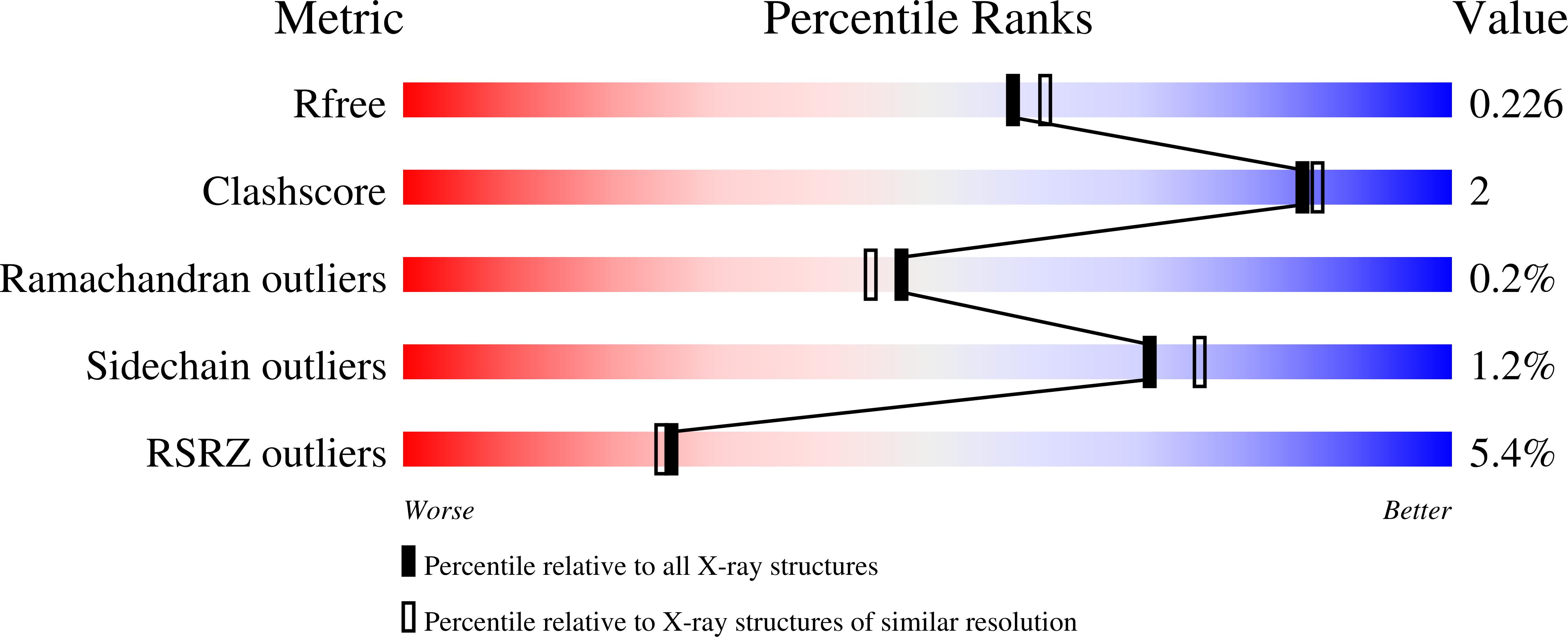
  • R-Value Free: 0.226 
  • R-Value Work: 0.181 
  • R-Value Observed: 0.184 

Starting Model: experimental
View more details

wwPDB Validation   3D Report Full Report


Ligand Structure Quality Assessment 


This is version 1.3 of the entry. See complete history


Literature

Determinants of the Inhibition of DprE1 and CYP2C9 by Antitubercular Thiophenes.

Liu, R.Lyu, X.Batt, S.M.Hsu, M.H.Harbut, M.B.Vilcheze, C.Cheng, B.Ajayi, K.Yang, B.Yang, Y.Guo, H.Lin, C.Gan, F.Wang, C.Franzblau, S.G.Jacobs, W.R.Besra, G.S.Johnson, E.F.Petrassi, M.Chatterjee, A.K.Futterer, K.Wang, F.

(2017) Angew Chem Int Ed Engl 56: 13011-13015

  • DOI: https://doi.org/10.1002/anie.201707324
  • Primary Citation of Related Structures:  
    5OEL, 5OEP, 5OEQ, 5W0C

  • PubMed Abstract: 

    Mycobacterium tuberculosis (Mtb) DprE1, an essential isomerase for the biosynthesis of the mycobacterial cell wall, is a validated target for tuberculosis (TB) drug development. Here we report the X-ray crystal structures of DprE1 and the DprE1 resistant mutant (Y314C) in complexes with TCA1 derivatives to elucidate the molecular basis of their inhibitory activities and an unconventional resistance mechanism, which enabled us to optimize the potency of the analogs. The selected lead compound showed excellent in vitro and in vivo activities, and low risk of toxicity profile except for the inhibition of CYP2C9. A crystal structure of CYP2C9 in complex with a TCA1 analog revealed the similar interaction patterns to the DprE1-TCA1 complex. Guided by the structures, an optimized molecule was generated with differential inhibitory activities against DprE1 and CYP2C9, which provides insights for development of a clinical candidate to treat TB.


  • Organizational Affiliation

    California Institute for Biomedical Research (Calibr), La Jolla, CA, 92037, USA.


Macromolecules
Find similar proteins by:  (by identity cutoff)  |  3D Structure
Entity ID: 1
MoleculeChains Sequence LengthOrganismDetailsImage
Cytochrome P450 2C9477Homo sapiensMutation(s): 0 
Gene Names: CYP2C9CYP2C10
EC: 1.14.13 (PDB Primary Data), 1.14.13.80 (PDB Primary Data), 1.14.13.48 (PDB Primary Data), 1.14.13.49 (PDB Primary Data), 1.14.99.38 (PDB Primary Data), 1.14.14.1 (UniProt), 1.14.14.53 (UniProt), 1.14.14.52 (UniProt), 1.14.14.51 (UniProt)
Membrane Entity: Yes 
UniProt & NIH Common Fund Data Resources
Find proteins for P11712 (Homo sapiens)
Explore P11712 
Go to UniProtKB:  P11712
PHAROS:  P11712
GTEx:  ENSG00000138109 
Entity Groups  
Sequence Clusters30% Identity50% Identity70% Identity90% Identity95% Identity100% Identity
UniProt GroupP11712
Sequence Annotations
Expand
  • Reference Sequence
Small Molecules
Binding Affinity Annotations 
IDSourceBinding Affinity
9W6 BindingDB:  5W0C IC50: 100 (nM) from 1 assay(s)
Experimental Data & Validation

Experimental Data

  • Method: X-RAY DIFFRACTION
  • Resolution: 2.00 Å
  • R-Value Free: 0.226 
  • R-Value Work: 0.181 
  • R-Value Observed: 0.184 
  • Space Group: H 3
Unit Cell:
Length ( Å )Angle ( ˚ )
a = 91.394α = 90
b = 91.394β = 90
c = 169.225γ = 120
Software Package:
Software NamePurpose
PHENIXrefinement
XDSdata reduction
Aimlessdata scaling
PHASERphasing

Structure Validation

View Full Validation Report


Ligand Structure Quality Assessment 


Entry History & Funding Information

Deposition Data


Funding OrganizationLocationGrant Number
National Institutes of Health/National Institute of General Medical Sciences (NIH/NIGMS)United StatesGM031001

Revision History  (Full details and data files)

  • Version 1.0: 2017-09-13
    Type: Initial release
  • Version 1.1: 2017-10-18
    Changes: Database references
  • Version 1.2: 2020-01-01
    Changes: Author supporting evidence
  • Version 1.3: 2023-10-04
    Changes: Data collection, Database references, Refinement description