5WI6 | pdb_00005wi6

Human beta-1 tryptase mutant Ile99Cys


Experimental Data Snapshot

  • Method: X-RAY DIFFRACTION
  • Resolution: 2.72 Å
  • R-Value Free: 
    0.227 (Depositor), 0.238 (DCC) 
  • R-Value Work: 
    0.193 (Depositor), 0.201 (DCC) 
  • R-Value Observed: 
    0.194 (Depositor) 

Starting Model: experimental
View more details

wwPDB Validation 3D Report Full Report

Validation slider image for 5WI6

Ligand Structure Quality Assessment 


This is version 1.5 of the entry. See complete history

Literature

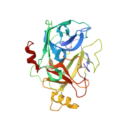
Dual functionality of beta-tryptase protomers as both proteases and cofactors in the active tetramer.

Maun, H.R.Liu, P.S.Franke, Y.Eigenbrot, C.Forrest, W.F.Schwartz, L.B.Lazarus, R.A.

(2018) J Biological Chem 293: 9614-9628

  • DOI: https://doi.org/10.1074/jbc.M117.812016
  • Primary Citation Related Structures: 
    5WI6

  • PubMed Abstract: 

    Human β-tryptase, a tetrameric trypsin-like serine protease, is an important mediator of the allergic inflammatory responses in asthma. During acute hypersensitivity reactions, mast cells degranulate, releasing active tetramer as a complex with proteoglycans. Extensive efforts have focused on developing therapeutic β-tryptase inhibitors, but its unique activation mechanism is less well-explored. Tryptase is active only after proteolytic removal of the pro-domain followed by tetramer formation via two distinct symmetry-related interfaces. We show that the cleaved I16G mutant cannot tetramerize, likely due to impaired insertion of its N terminus into its "activation pocket," indicating allosteric linkage at multiple sites on each protomer. We engineered cysteines into each of the two distinct interfaces (Y75C for small or I99C for large) to assess the activity of each tetramer and disulfide-locked dimer. Using size-exclusion chromatography and enzymatic assays, we demonstrate that the two large tetramer interfaces regulate enzymatic activity, elucidating the importance of this protein-protein interaction for allosteric regulation. Notably, the I99C large interface dimer is active, even in the absence of heparin. We show that a monomeric β-tryptase mutant (I99C*/Y75A/Y37bA, where C* is cysteinylated Cys-99) cannot form a dimer or tetramer, yet it is active but only in the presence of heparin. Thus heparin both stabilizes the tetramer and allosterically conditions the active site. We hypothesize that each β-tryptase protomer in the tetramer has two distinct roles, acting both as a protease and as a cofactor for its neighboring protomer, to allosterically regulate enzymatic activity, providing a rationale for direct correlation of tetramer stability with proteolytic activity.


  • Organizational Affiliation
    • From the Departments of Early Discovery Biochemistry.

Macromolecule Content 

  • Total Structure Weight: 112.12 kDa 
  • Atom Count: 7,828 
  • Modeled Residue Count: 972 
  • Deposited Residue Count: 980 
  • Unique protein chains: 1

Macromolecules

Find similar proteins by:|  3D Structure
Entity ID: 1
MoleculeChains  Sequence LengthOrganismDetailsImage
Tryptase alpha/beta-1
A, B, C, D
245Homo sapiensMutation(s): 1 
Gene Names: TPSAB1TPS1TPS2TPSB1
EC: 3.4.21.59
UniProt & NIH Common Fund Data Resources
Find proteins for Q15661 (Homo sapiens)
Explore Q15661 
Go to UniProtKB:  Q15661
PHAROS:  Q15661
GTEx:  ENSG00000172236 
Entity Groups
Sequence Clusters30% Identity50% Identity70% Identity90% Identity95% Identity100% Identity
UniProt GroupQ15661
Sequence Annotations
Expand
Reference Sequence

Small Molecules

Ligands 2 Unique
IDChains Name / Formula / InChI Key2D Diagram3D Interactions
0GJ

Query on 0GJ


Download:Ideal Coordinates CCD File
F [auth A],
H [auth B],
K [auth C],
O [auth D]
L-alpha-glutamyl-N-{(1S)-4-{[amino(iminio)methyl]amino}-1-[(1S)-2-chloro-1-hydroxyethyl]butyl}glycinamide
C14 H28 Cl N6 O5
XELWNHKFCNMWQO-LPEHRKFASA-O
SO4

Query on SO4


Download:Ideal Coordinates CCD File
E [auth A]
G [auth B]
I [auth C]
J [auth C]
L [auth D]
E [auth A],
G [auth B],
I [auth C],
J [auth C],
L [auth D],
M [auth D],
N [auth D]
SULFATE ION
O4 S
QAOWNCQODCNURD-UHFFFAOYSA-L

Experimental Data & Validation

Experimental Data

  • Method: X-RAY DIFFRACTION
  • Resolution: 2.72 Å
  • R-Value Free:  0.227 (Depositor), 0.238 (DCC) 
  • R-Value Work:  0.193 (Depositor), 0.201 (DCC) 
  • R-Value Observed: 0.194 (Depositor) 
Space Group: P 32 2 1
Unit Cell:
Length ( Å )Angle ( ˚ )
a = 96.743α = 90
b = 96.743β = 90
c = 238.553γ = 120
Software Package:
Software NamePurpose
BUSTERrefinement
XDSdata reduction
Aimlessdata scaling
PHASERphasing

Structure Validation

View Full Validation Report


Ligand Structure Quality Assessment 


Entry History 

Deposition Data

Revision History  (Full details and data files)

  • Version 1.0: 2018-04-25
    Type: Initial release
  • Version 1.1: 2018-05-02
    Changes: Data collection, Database references
  • Version 1.2: 2018-05-16
    Changes: Data collection, Structure summary
  • Version 1.3: 2018-07-04
    Changes: Data collection, Database references
  • Version 1.4: 2023-10-04
    Changes: Data collection, Database references, Refinement description
  • Version 1.5: 2024-10-16
    Changes: Structure summary