5WQ0 | pdb_00005wq0

Receiver domain of Spo0A from Paenisporosarcina sp. TG-14


Experimental Data Snapshot

  • Method: X-RAY DIFFRACTION
  • Resolution: 2.60 Å
  • R-Value Free: 
    0.243 (Depositor), 0.246 (DCC) 
  • R-Value Work: 
    0.187 (Depositor), 0.191 (DCC) 
  • R-Value Observed: 
    0.190 (Depositor) 

wwPDB Validation 3D Report Full Report

Validation slider image for 5WQ0

This is version 1.2 of the entry. See complete history

Literature

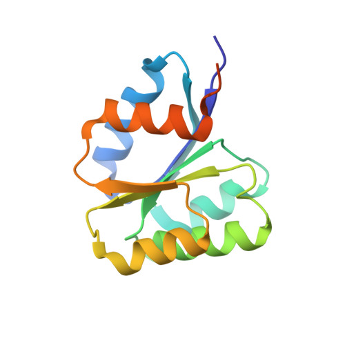
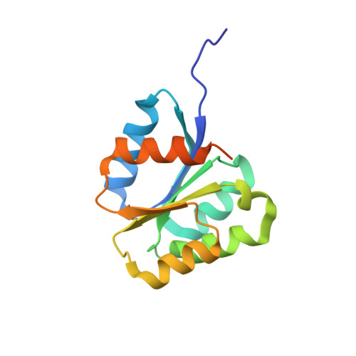
Crystal structure of the inactive state of the receiver domain of Spo0A from Paenisporosarcina sp. TG-14, a psychrophilic bacterium isolated from an Antarctic glacier

Lee, C.W.Park, S.H.Lee, S.G.Shin, S.C.Han, S.J.Kim, H.W.Park, H.H.Kim, S.Kim, H.J.Park, H.Park, H.Lee, J.H.

(2017) J Microbiol 55: 464-474

  • DOI: https://doi.org/10.1007/s12275-017-6599-9
  • Primary Citation Related Structures: 
    5WQ0

  • PubMed Abstract: 

    The two-component phosphorelay system is the most prevalent mechanism for sensing and transducing environmental signals in bacteria. Spore formation, which relies on the two-component phosphorelay system, enables the long-term survival of the glacial bacterium Paenisporosarcina sp. TG-14 in the extreme cold environment. Spo0A is a key response regulator of the phosphorelay system in the early stage of spore formation. The protein is composed of a regulatory N-terminal phospho-receiver domain and a DNA-binding C-terminal activator domain. We solved the three-dimensional structure of the unphosphorylated (inactive) form of the receiver domain of Spo0A (PaSpo0A-R) from Paenisporosarcina sp. TG-14. A structural comparison with phosphorylated (active form) Spo0A from Bacillus stearothermophilus (BsSpo0A) showed minor notable differences. A molecular dynamics study of a model of the active form and the crystal structures revealed significant differences in the α4 helix and the preceding loop region where phosphorylation occurs. Although an oligomerization study of PaSpo0A-R by analytical ultracentrifugation (AUC) has shown that the protein is in a monomeric state in solution, both crosslinking and crystal-packing analyses indicate the possibility of weak dimer formation by a previously undocumented mechanism. Collectively, these observations provide insight into the mechanism of phosphorylation-dependent activation unique to Spo0A.


  • Organizational Affiliation
    • Unit of Polar Genomics, Korea Polar Research Institute, Incheon, 21990, Republic of Korea.

Macromolecule Content 

  • Total Structure Weight: 113.41 kDa 
  • Atom Count: 6,893 
  • Modeled Residue Count: 873 
  • Deposited Residue Count: 1,012 
  • Unique protein chains: 2

Macromolecules

Find similar proteins by:|  3D Structure
Entity ID: 1
MoleculeChains  Sequence LengthOrganismDetailsImage
Stage 0 sporulation protein144Paenisporosarcina sp. TG-14Mutation(s): 0 
UniProt
Find proteins for A0ACD6BAT0 (Paenisporosarcina sp. TG-14)
Explore A0ACD6BAT0 
Go to UniProtKB:  A0ACD6BAT0
Entity Groups
Sequence Clusters30% Identity50% Identity70% Identity90% Identity95% Identity100% Identity
UniProt GroupA0ACD6BAT0
Sequence Annotations
Expand
Reference Sequence
Find similar proteins by:|  3D Structure
Entity ID: 2
MoleculeChains  Sequence LengthOrganismDetailsImage
Stage 0 sporulation protein148Paenisporosarcina sp. TG-14Mutation(s): 0 
UniProt
Find proteins for A0ACD6BAX7 (Paenisporosarcina sp. TG-14)
Explore A0ACD6BAX7 
Go to UniProtKB:  A0ACD6BAX7
Entity Groups
Sequence Clusters30% Identity50% Identity70% Identity90% Identity95% Identity100% Identity
UniProt GroupA0ACD6BAX7
Sequence Annotations
Expand
Reference Sequence

Experimental Data & Validation

Experimental Data

  • Method: X-RAY DIFFRACTION
  • Resolution: 2.60 Å
  • R-Value Free:  0.243 (Depositor), 0.246 (DCC) 
  • R-Value Work:  0.187 (Depositor), 0.191 (DCC) 
  • R-Value Observed: 0.190 (Depositor) 
Space Group: P 21 21 21
Unit Cell:
Length ( Å )Angle ( ˚ )
a = 56.969α = 90
b = 72.233β = 90
c = 189.65γ = 90
Software Package:
Software NamePurpose
PHENIXrefinement
HKL-2000data scaling
HKL-2000data processing
Cootmodel building

Structure Validation

View Full Validation Report


Entry History 

Deposition Data

Revision History  (Full details and data files)

  • Version 1.0: 2017-03-22
    Type: Initial release
  • Version 1.1: 2017-06-07
    Changes: Database references
  • Version 1.2: 2024-03-20
    Changes: Data collection, Database references, Derived calculations