5YB6

L-Amino acid oxidase/monooxygenase from Pseudomonas sp. AIU 813 - L-lysine complex


Experimental Data Snapshot

  • Method: X-RAY DIFFRACTION
  • Resolution: 2.10 Å
  • R-Value Free: 
    0.283 (Depositor), 0.290 (DCC) 
  • R-Value Work: 
    0.234 (Depositor), 0.240 (DCC) 
  • R-Value Observed: 
    0.237 (Depositor) 

Starting Model: experimental
View more details

wwPDB Validation   3D Report Full Report


Ligand Structure Quality Assessment 

Created with Raphaël 2.3.0Worse 01 BetterLigand structure goodness of fit to experimental dataBest fitted FADClick on this verticalbar to view details

This is version 1.2 of the entry. See complete history


Literature

Ligand complex structures of l-amino acid oxidase/monooxygenase from

Im, D.Matsui, D.Arakawa, T.Isobe, K.Asano, Y.Fushinobu, S.

(2018) FEBS Open Bio 8: 314-324

  • DOI: https://doi.org/10.1002/2211-5463.12387
  • Primary Citation of Related Structures:  
    5YB6, 5YB7, 5YB8

  • PubMed Abstract: 

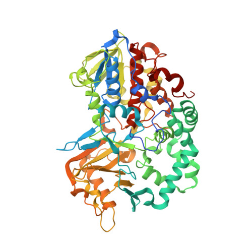
    l-Amino acid oxidase/monooxygenase from Pseudomonas sp. AIU 813 (l-AAO/MOG) catalyzes both the oxidative deamination and oxidative decarboxylation of the α-group of l-Lys to produce a keto acid and amide, respectively. l-AAO/MOG exhibits limited specificity for l-amino acid substrates with a basic side chain. We previously determined its ligand-free crystal structure and identified a key residue for maintaining the dual activities. Here, we determined the structures of l-AAO/MOG complexed with l-Lys, l-ornithine, and l-Arg and revealed its substrate recognition. Asp238 is located at the ceiling of a long hydrophobic pocket and forms a strong interaction with the terminal, positively charged group of the substrates. A mutational analysis on the D238A mutant indicated that the interaction is critical for substrate binding but not for catalytic control between the oxidase/monooxygenase activities. The catalytic activities of the D238E mutant unexpectedly increased, while the D238F mutant exhibited altered substrate specificity to long hydrophobic substrates. In the ligand-free structure, there are two channels connecting the active site and solvent, and a short region located at the dimer interface is disordered. In the l-Lys complex structure, a loop region is displaced to plug the channels. Moreover, the disordered region in the ligand-free structure forms a short helix in the substrate complex structures and creates the second binding site for the substrate. It is assumed that the amino acid substrate enters the active site of l-AAO/MOG through this route. The atomic coordinates and structure factors (codes 5YB6, 5YB7, and 5YB8) have been deposited in the Protein Data Bank (http://wwpdb.org/). 1.4.3.2 (l-amino acid oxidase), 1.13.12.2 (lysine 2-monooxygenase).


  • Organizational Affiliation

    Department of Biotechnology The University of Tokyo Japan.


Macromolecules
Find similar proteins by:  (by identity cutoff)  |  3D Structure
Entity ID: 1
MoleculeChains Sequence LengthOrganismDetailsImage
L-amino acid oxidase/monooxygenase
A, B, C, D
580Pseudomonas sp. AIU 813Mutation(s): 0 
Gene Names: laaomog
EC: 1.13.12.3
UniProt
Find proteins for W6JQJ6 (Pseudomonas sp. AIU 813)
Explore W6JQJ6 
Go to UniProtKB:  W6JQJ6
Entity Groups  
Sequence Clusters30% Identity50% Identity70% Identity90% Identity95% Identity100% Identity
UniProt GroupW6JQJ6
Sequence Annotations
Expand
  • Reference Sequence
Small Molecules
Ligands 3 Unique
IDChains Name / Formula / InChI Key2D Diagram3D Interactions
FAD
Query on FAD

Download Ideal Coordinates CCD File 
E [auth A],
I [auth B],
M [auth C],
Q [auth D]
FLAVIN-ADENINE DINUCLEOTIDE
C27 H33 N9 O15 P2
VWWQXMAJTJZDQX-UYBVJOGSSA-N
PG6
Query on PG6

Download Ideal Coordinates CCD File 
F [auth A],
J [auth B],
N [auth C],
R [auth D]
1-(2-METHOXY-ETHOXY)-2-{2-[2-(2-METHOXY-ETHOXY]-ETHOXY}-ETHANE
C12 H26 O6
DMDPGPKXQDIQQG-UHFFFAOYSA-N
LYS
Query on LYS

Download Ideal Coordinates CCD File 
G [auth A]
H [auth A]
K [auth B]
L [auth B]
O [auth C]
LYSINE
C6 H15 N2 O2
KDXKERNSBIXSRK-YFKPBYRVSA-O
Experimental Data & Validation

Experimental Data

  • Method: X-RAY DIFFRACTION
  • Resolution: 2.10 Å
  • R-Value Free:  0.283 (Depositor), 0.290 (DCC) 
  • R-Value Work:  0.234 (Depositor), 0.240 (DCC) 
  • R-Value Observed: 0.237 (Depositor) 
Space Group: P 1 21 1
Unit Cell:
Length ( Å )Angle ( ˚ )
a = 97.071α = 90
b = 132.293β = 108.67
c = 100.963γ = 90
Software Package:
Software NamePurpose
HKL-2000data processing
HKL-2000data scaling
MOLREPphasing
REFMACrefinement
PDB_EXTRACTdata extraction

Structure Validation

View Full Validation Report


Ligand Structure Quality Assessment 

Created with Raphaël 2.3.0Worse 01 BetterLigand structure goodness of fit to experimental dataBest fitted FADClick on this verticalbar to view details

Entry History 

Deposition Data

Revision History  (Full details and data files)

  • Version 1.0: 2018-02-07
    Type: Initial release
  • Version 1.1: 2018-03-21
    Changes: Database references
  • Version 1.2: 2023-11-22
    Changes: Data collection, Database references, Refinement description