6BL5 | pdb_00006bl5

Head decoration protein from the hyperthermophilic phage P74-26


Experimental Data Snapshot

  • Method: X-RAY DIFFRACTION
  • Resolution: 1.69 Å
  • R-Value Free: 
    0.213 (Depositor), 0.215 (DCC) 
  • R-Value Work: 
    0.179 (Depositor), 0.180 (DCC) 
  • R-Value Observed: 
    0.182 (Depositor) 

wwPDB Validation 3D Report Full Report

Validation slider image for 6BL5

This is version 1.3 of the entry. See complete history

Literature

A Hyperthermophilic Phage Decoration Protein Suggests Common Evolutionary Origin with Herpesvirus Triplex Proteins and an Anti-CRISPR Protein.

Stone, N.P.Hilbert, B.J.Hidalgo, D.Halloran, K.T.Lee, J.Sontheimer, E.J.Kelch, B.A.

(2018) Structure 26: 936-947.e3

  • DOI: https://doi.org/10.1016/j.str.2018.04.008
  • Primary Citation Related Structures: 
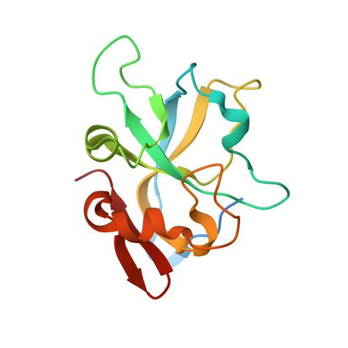
    6BL5

  • PubMed Abstract: 

    Virus capsids are protein shells that protect the viral genome from environmental assaults, while maintaining the high internal pressure of the tightly packaged genome. To elucidate how capsids maintain stability under harsh conditions, we investigated the capsid components of the hyperthermophilic phage P74-26. We determined the structure of capsid protein gp87 and show that it has the same fold as decoration proteins in many other phages, despite lacking significant sequence homology. We also find that gp87 is significantly more stable than mesophilic homologs. Our analysis of the gp87 structure reveals that the core "β tulip" domain is conserved in trimeric capsid components across numerous double-stranded DNA viruses, including Herpesviruses. Moreover, this β barrel domain is found in anti-CRISPR protein AcrIIC1, suggesting a mechanism for the evolution of this Cas9 inhibitor. Our work illustrates the principles for increased stability of gp87, and extends the evolutionary reach of the β tulip domain.


  • Organizational Affiliation
    • Department of Biochemistry and Molecular Pharmacology, University of Massachusetts Medical School, Worcester, MA 01655, USA.

Macromolecule Content 

  • Total Structure Weight: 16.25 kDa 
  • Atom Count: 1,106 
  • Modeled Residue Count: 129 
  • Deposited Residue Count: 145 
  • Unique protein chains: 1

Macromolecules

Find similar proteins by:|  3D Structure
Entity ID: 1
MoleculeChains  Sequence LengthOrganismDetailsImage
Head decoration protein145Oshimavirus P7426Mutation(s): 0 
Gene Names: P74p87
UniProt
Find proteins for A7XXR5 (Thermus phage P74-26)
Explore A7XXR5 
Go to UniProtKB:  A7XXR5
Entity Groups
Sequence Clusters30% Identity50% Identity70% Identity90% Identity95% Identity100% Identity
UniProt GroupA7XXR5
Sequence Annotations
Expand
Reference Sequence

Experimental Data & Validation

Experimental Data

  • Method: X-RAY DIFFRACTION
  • Resolution: 1.69 Å
  • R-Value Free:  0.213 (Depositor), 0.215 (DCC) 
  • R-Value Work:  0.179 (Depositor), 0.180 (DCC) 
  • R-Value Observed: 0.182 (Depositor) 
Space Group: P 63
Unit Cell:
Length ( Å )Angle ( ˚ )
a = 89.516α = 90
b = 89.516β = 90
c = 31.39γ = 120
Software Package:
Software NamePurpose
PHENIXrefinement
HKL-3000data collection
HKL-3000data scaling
Cootmodel building
AutoSolphasing
HKL-3000data reduction

Structure Validation

View Full Validation Report


Entry History 

& Funding Information

Deposition Data


Funding OrganizationLocationGrant Number
The Pew Charitable TrustsUnited States--

Revision History  (Full details and data files)

  • Version 1.0: 2018-02-21
    Type: Initial release
  • Version 1.1: 2018-06-06
    Changes: Data collection, Database references
  • Version 1.2: 2018-07-18
    Changes: Data collection, Database references
  • Version 1.3: 2024-03-13
    Changes: Data collection, Database references