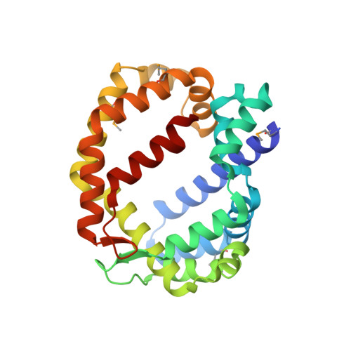
6BR8 | pdb_00006br8

Structure of A6 reveals a novel lipid transporter


Experimental Data Snapshot

  • Method: X-RAY DIFFRACTION
  • Resolution: 2.30 Å
  • R-Value Free: 
    0.289 (Depositor), 0.289 (DCC) 
  • R-Value Work: 
    0.228 (Depositor), 0.236 (DCC) 
  • R-Value Observed: 
    0.233 (Depositor) 

wwPDB Validation   3D Report Full Report


Ligand Structure Quality Assessment 

Created with Raphaël 2.3.0Worse 01 BetterLigand structure goodness of fit to experimental dataBest fitted PGVClick on this verticalbar to view detailsBest fitted 6OUClick on this verticalbar to view details

This is version 1.5 of the entry. See complete history


Literature

Structure of a lipid-bound viral membrane assembly protein reveals a modality for enclosing the lipid bilayer.

Pathak, P.K.Peng, S.Meng, X.Han, Y.Zhang, B.Zhang, F.Xiang, Y.Deng, J.

(2018) Proc Natl Acad Sci U S A 115: 7028-7032

  • DOI: https://doi.org/10.1073/pnas.1805855115
  • Primary Citation of Related Structures:  
    6BR8, 6BR9, 6CB6, 6CB7

  • PubMed Abstract: 

    Cellular membranes are maintained as closed compartments, broken up only transiently during membrane reorganization or lipid transportation. However, open-ended membranes, likely derived from scissions of the endoplasmic reticulum, persist in vaccinia virus-infected cells during the assembly of the viral envelope. A group of viral membrane assembly proteins (VMAPs) were identified as essential for this process. To understand the mechanism of VMAPs, we determined the 2.2-Å crystal structure of the largest member, named A6, which is a soluble protein with two distinct domains. The structure of A6 displays a novel protein fold composed mainly of alpha helices. The larger C-terminal domain forms a unique cage that encloses multiple glycerophospholipids with a lipid bilayer-like configuration. The smaller N-terminal domain does not bind lipid but negatively affects lipid binding by A6. Mutations of key hydrophobic residues lining the lipid-binding cage disrupt lipid binding and abolish viral replication. Our results reveal a protein modality for enclosing the lipid bilayer and provide molecular insight into a viral machinery involved in generating and/or stabilizing open-ended membranes.


  • Organizational Affiliation

    Department of Biochemistry and Molecular Biology, Oklahoma State University, Stillwater, OK 74078.


Macromolecules
Find similar proteins by:  (by identity cutoff)  |  3D Structure
Entity ID: 1
MoleculeChains Sequence LengthOrganismDetailsImage
Protein A6 homolog
A, B
252Fowlpox virus strain NVSLMutation(s): 0 
Gene Names: FPV170
UniProt
Find proteins for Q9J563 (Fowlpox virus (strain NVSL))
Explore Q9J563 
Go to UniProtKB:  Q9J563
Entity Groups  
Sequence Clusters30% Identity50% Identity70% Identity90% Identity95% Identity100% Identity
UniProt GroupQ9J563
Sequence Annotations
Expand
  • Reference Sequence
Small Molecules
Ligands 2 Unique
IDChains Name / Formula / InChI Key2D Diagram3D Interactions
PGV
Query on PGV

Download Ideal Coordinates CCD File 
F [auth A],
K [auth B]
(1R)-2-{[{[(2S)-2,3-DIHYDROXYPROPYL]OXY}(HYDROXY)PHOSPHORYL]OXY}-1-[(PALMITOYLOXY)METHYL]ETHYL (11E)-OCTADEC-11-ENOATE
C40 H77 O10 P
ADYWCMPUNIVOEA-GPJPVTGXSA-N
6OU
Query on 6OU

Download Ideal Coordinates CCD File 
C [auth A]
D [auth A]
E [auth A]
G [auth A]
H [auth B]
[(2~{R})-1-[2-azanylethoxy(oxidanyl)phosphoryl]oxy-3-hexadecanoyloxy-propan-2-yl] (~{Z})-octadec-9-enoate
C39 H76 N O8 P
FHQVHHIBKUMWTI-OTMQOFQLSA-N
Modified Residues  1 Unique
IDChains TypeFormula2D DiagramParent
MSE
Query on MSE
A, B
L-PEPTIDE LINKINGC5 H11 N O2 SeMET
Experimental Data & Validation

Experimental Data

  • Method: X-RAY DIFFRACTION
  • Resolution: 2.30 Å
  • R-Value Free:  0.289 (Depositor), 0.289 (DCC) 
  • R-Value Work:  0.228 (Depositor), 0.236 (DCC) 
  • R-Value Observed: 0.233 (Depositor) 
Space Group: P 21 21 21
Unit Cell:
Length ( Å )Angle ( ˚ )
a = 54.909α = 90
b = 75.163β = 90
c = 148.38γ = 90
Software Package:
Software NamePurpose
PHENIXrefinement
HKL-3000data reduction
HKL-3000data scaling
PHENIXphasing

Structure Validation

View Full Validation Report


Ligand Structure Quality Assessment 

Created with Raphaël 2.3.0Worse 01 BetterLigand structure goodness of fit to experimental dataBest fitted PGVClick on this verticalbar to view detailsBest fitted 6OUClick on this verticalbar to view details

Entry History & Funding Information

Deposition Data


Funding OrganizationLocationGrant Number
National Institutes of Health/National Institute Of Allergy and Infectious Diseases (NIH/NIAID)United StatesAI 133589

Revision History  (Full details and data files)

  • Version 1.0: 2018-06-20
    Type: Initial release
  • Version 1.1: 2018-07-04
    Changes: Data collection, Database references
  • Version 1.2: 2018-07-18
    Changes: Data collection, Database references
  • Version 1.3: 2019-02-20
    Changes: Author supporting evidence, Data collection
  • Version 1.4: 2019-12-11
    Changes: Author supporting evidence
  • Version 1.5: 2024-11-20
    Changes: Advisory, Data collection, Database references, Structure summary