6CE0 | pdb_00006ce0

Crystal structure of a HIV-1 clade B tier-3 isolate H078.14 UFO-BG Env trimer in complex with broadly neutralizing Fabs PGT124 and 35O22 at 4.6 Angstrom


Experimental Data Snapshot

  • Method: X-RAY DIFFRACTION
  • Resolution: 4.60 Å
  • R-Value Free: 
    0.341 (Depositor), 0.345 (DCC) 
  • R-Value Work: 
    0.313 (Depositor), 0.314 (DCC) 
  • R-Value Observed: 
    0.314 (Depositor) 

Starting Models: experimental
View more details

wwPDB Validation 3D Report Full Report

Validation slider image for 6CE0

Ligand Structure Quality Assessment 


This is version 3.0 of the entry. See complete history

Literature

HIV-1 vaccine design through minimizing envelope metastability.

He, L.Kumar, S.Allen, J.D.Huang, D.Lin, X.Mann, C.J.Saye-Francisco, K.L.Copps, J.Sarkar, A.Blizard, G.S.Ozorowski, G.Sok, D.Crispin, M.Ward, A.B.Nemazee, D.Burton, D.R.Wilson, I.A.Zhu, J.

(2018) Sci Adv 4: eaau6769-eaau6769

  • DOI: https://doi.org/10.1126/sciadv.aau6769
  • Primary Citation Related Structures: 
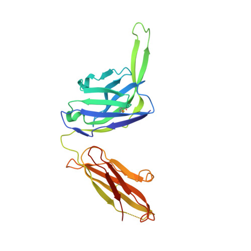
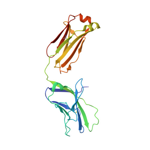
    6CE0

  • PubMed Abstract: 

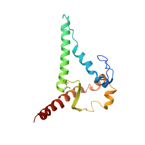
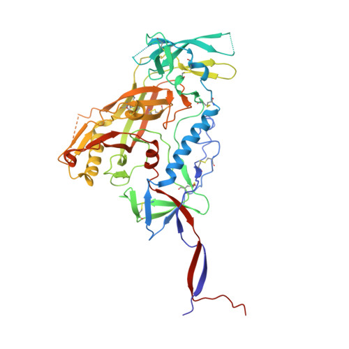
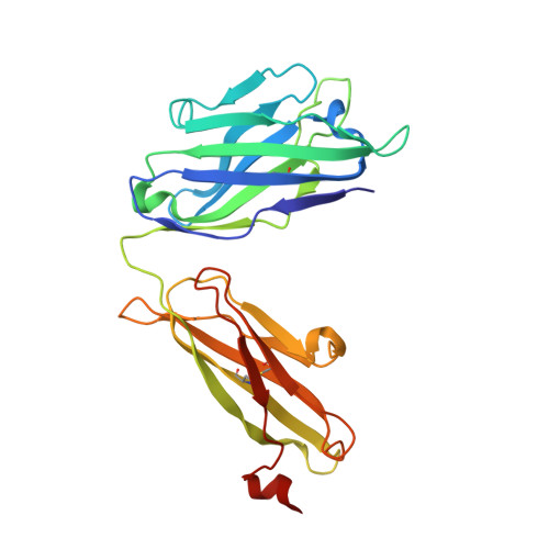
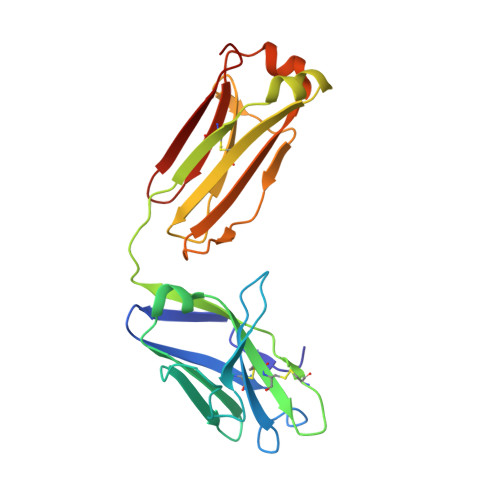
    Overcoming envelope metastability is crucial to trimer-based HIV-1 vaccine design. Here, we present a coherent vaccine strategy by minimizing metastability. For 10 strains across five clades, we demonstrate that the gp41 ectodomain (gp41 ECTO ) is the main source of envelope metastability by replacing wild-type gp41 ECTO with BG505 gp41 ECTO of the uncleaved prefusion-optimized (UFO) design. These gp41 ECTO -swapped trimers can be produced in CHO cells with high yield and high purity. The crystal structure of a gp41 ECTO -swapped trimer elucidates how a neutralization-resistant tier 3 virus evades antibody recognition of the V2 apex. UFO trimers of transmitted/founder viruses and UFO trimers containing a consensus-based ancestral gp41 ECTO suggest an evolutionary root of metastability. The gp41 ECTO -stabilized trimers can be readily displayed on 24- and 60-meric nanoparticles, with incorporation of additional T cell help illustrated for a hyperstable 60-mer, I3-01. In mice and rabbits, these gp140 nanoparticles induced tier 2 neutralizing antibody responses more effectively than soluble trimers.


  • Organizational Affiliation
    • Department of Immunology and Microbiology, The Scripps Research Institute, La Jolla, CA 92037, USA.

Macromolecule Content 

  • Total Structure Weight: 176.94 kDa 
  • Atom Count: 11,946 
  • Modeled Residue Count: 1,473 
  • Deposited Residue Count: 1,536 
  • Unique protein chains: 6

Macromolecules

Find similar proteins by:|  3D Structure
Entity ID: 1
MoleculeChains  Sequence LengthOrganismDetailsImage
Envelope glycoprotein gp160A [auth B]140Human immunodeficiency virus 1Mutation(s): 1 
Gene Names: env
UniProt
Find proteins for Q2N0S5 (Human immunodeficiency virus type 1)
Explore Q2N0S5 
Go to UniProtKB:  Q2N0S5
Find proteins for Q2N0T3 (Human immunodeficiency virus type 1)
Explore Q2N0T3 
Go to UniProtKB:  Q2N0T3
Entity Groups
Sequence Clusters30% Identity50% Identity70% Identity90% Identity95% Identity100% Identity
UniProt GroupsQ2N0T3Q2N0S5
Glycosylation
Glycosylation Sites: 1
Sequence Annotations
Expand
Reference Sequence
Find similar proteins by:|  3D Structure
Entity ID: 2
MoleculeChains  Sequence LengthOrganismDetailsImage
Envelope glycoprotein gp160B [auth G]487Human immunodeficiency virus 1Mutation(s): 1 
Gene Names: env
UniProt
Find proteins for A4ZPX1 (Human immunodeficiency virus type 1)
Explore A4ZPX1 
Go to UniProtKB:  A4ZPX1
Entity Groups
Sequence Clusters30% Identity50% Identity70% Identity90% Identity95% Identity100% Identity
UniProt GroupA4ZPX1
Glycosylation
Glycosylation Sites: 21
Sequence Annotations
Expand
Reference Sequence
Find similar proteins by:|  3D Structure
Entity ID: 3
MoleculeChains  Sequence LengthOrganismDetailsImage
35O22 Heavy chainC [auth D]243Homo sapiensMutation(s): 0 
Find similar proteins by:|  3D Structure
Entity ID: 4
MoleculeChains  Sequence LengthOrganismDetailsImage
35O22 Light chainD [auth E]216Homo sapiensMutation(s): 0 
Find similar proteins by:|  3D Structure
Entity ID: 5
MoleculeChains  Sequence LengthOrganismDetailsImage
PGT124 Heavy chainE [auth H]236Homo sapiensMutation(s): 0 
Find similar proteins by:|  3D Structure
Entity ID: 6
MoleculeChains  Sequence LengthOrganismDetailsImage
PGT124 Light chainF [auth L]214Homo sapiensMutation(s): 0 

Oligosaccharides

Help  
Entity ID: 7
MoleculeChains Length2D Diagram GlycosylationD Interactions
2-acetamido-2-deoxy-beta-D-glucopyranose-(1-4)-2-acetamido-2-deoxy-beta-D-glucopyranoseG [auth A],
H [auth C],
K [auth J]
2N-Glycosylation
Glycosylation Resources
GlyTouCan: G42666HT
GlyCosmos: G42666HT
GlyGen: G42666HT
Entity ID: 8
MoleculeChains Length2D Diagram GlycosylationD Interactions
beta-D-mannopyranose-(1-4)-2-acetamido-2-deoxy-beta-D-glucopyranose-(1-4)-2-acetamido-2-deoxy-beta-D-glucopyranoseI [auth F],
L [auth K]
3N-Glycosylation
Glycosylation Resources
GlyTouCan: G15407YE
GlyCosmos: G15407YE
GlyGen: G15407YE
Entity ID: 9
MoleculeChains Length2D Diagram GlycosylationD Interactions
alpha-D-mannopyranose-(1-3)-[alpha-D-mannopyranose-(1-6)]beta-D-mannopyranose-(1-4)-2-acetamido-2-deoxy-beta-D-glucopyranose-(1-4)-2-acetamido-2-deoxy-beta-D-glucopyranoseJ [auth I],
N
5N-Glycosylation
Glycosylation Resources
GlyTouCan: G22768VO
GlyCosmos: G22768VO
GlyGen: G22768VO
Entity ID: 10
MoleculeChains Length2D Diagram GlycosylationD Interactions
alpha-D-mannopyranose-(1-3)-beta-D-mannopyranose-(1-4)-2-acetamido-2-deoxy-beta-D-glucopyranose-(1-4)-2-acetamido-2-deoxy-beta-D-glucopyranose
M
4N-Glycosylation
Glycosylation Resources
GlyTouCan: G81315DD
GlyCosmos: G81315DD
GlyGen: G81315DD
Entity ID: 11
MoleculeChains Length2D Diagram GlycosylationD Interactions
alpha-D-mannopyranose-(1-2)-alpha-D-mannopyranose-(1-3)-[alpha-D-mannopyranose-(1-3)-alpha-D-mannopyranose-(1-6)]beta-D-mannopyranose-(1-4)-2-acetamido-2-deoxy-beta-D-glucopyranose-(1-4)-2-acetamido-2-deoxy-beta-D-glucopyranose
O
7N-Glycosylation
Glycosylation Resources
GlyTouCan: G07617FP
GlyCosmos: G07617FP
GlyGen: G07617FP

Small Molecules

Ligands 1 Unique
IDChains Name / Formula / InChI Key2D Diagram3D Interactions
NAG

Query on NAG


Download:Ideal Coordinates CCD File
AA [auth G]
BA [auth G]
P [auth G]
Q [auth G]
R [auth G]
AA [auth G],
BA [auth G],
P [auth G],
Q [auth G],
R [auth G],
S [auth G],
T [auth G],
U [auth G],
V [auth G],
W [auth G],
X [auth G],
Y [auth G],
Z [auth G]
2-acetamido-2-deoxy-beta-D-glucopyranose
C8 H15 N O6
OVRNDRQMDRJTHS-FMDGEEDCSA-N

Experimental Data & Validation

Experimental Data

  • Method: X-RAY DIFFRACTION
  • Resolution: 4.60 Å
  • R-Value Free:  0.341 (Depositor), 0.345 (DCC) 
  • R-Value Work:  0.313 (Depositor), 0.314 (DCC) 
  • R-Value Observed: 0.314 (Depositor) 
Space Group: P 63
Unit Cell:
Length ( Å )Angle ( ˚ )
a = 127.351α = 90
b = 127.351β = 90
c = 316.35γ = 120
Software Package:
Software NamePurpose
PHENIXrefinement
HKL-2000data reduction
HKL-2000data scaling
PHASERphasing

Structure Validation

View Full Validation Report


Ligand Structure Quality Assessment 


Entry History 

Deposition Data

Revision History  (Full details and data files)

  • Version 1.0: 2018-12-05
    Type: Initial release
  • Version 2.0: 2020-07-29
    Type: Remediation
    Reason: Carbohydrate remediation
    Changes: Advisory, Atomic model, Data collection, Derived calculations, Structure summary
  • Version 2.1: 2023-10-04
    Changes: Data collection, Database references, Derived calculations, Refinement description, Structure summary
  • Version 3.0: 2024-12-25
    Changes: Advisory, Atomic model, Data collection, Derived calculations, Structure summary