6CI0 | pdb_00006ci0

Catalytic core subunits (I and II) of cytochrome C oxidase from Rhodobacter sphaeroides with E101A (II) mutation


Experimental Data Snapshot

  • Method: X-RAY DIFFRACTION
  • Resolution: 2.40 Å
  • R-Value Free: 
    0.234 (Depositor), 0.232 (DCC) 
  • R-Value Work: 
    0.199 (Depositor), 0.200 (DCC) 
  • R-Value Observed: 
    0.200 (Depositor) 

Starting Model: experimental
View more details

wwPDB Validation 3D Report Full Report

Validation slider image for 6CI0

Ligand Structure Quality Assessment 


This is version 2.2 of the entry. See complete history

Literature

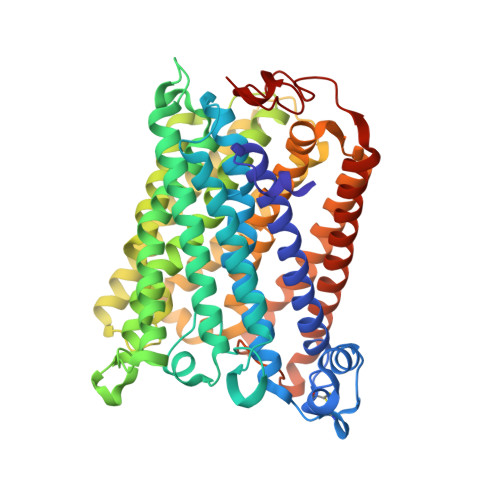
The K-path entrance in cytochrome c oxidase is defined by mutation of E101 and controlled by an adjacent ligand binding domain.

Hiser, C.Liu, J.Ferguson-Miller, S.

(2018) Biochim Biophys Acta 1859: 725-733

  • DOI: https://doi.org/10.1016/j.bbabio.2018.03.017
  • Primary Citation Related Structures: 
    6CI0

  • PubMed Abstract: 

    Three mutant forms of Rhodobacter sphaeroides cytochrome c oxidase (RsCcO) were created to test for multiple K-path entry sites (E101W), the existence of an "upper ligand site" (M350 W), and the nature and binding specificity of the "lower ligand site" (P315W/E101A) in the region of a crystallographically-defined deoxycholate at the K-path entrance. The effects of inhibitory and stimulatory detergents (dodecyl maltoside and Tween20) on these mutants are presented, as well as competition with other ligands, including the potentially physiologically relevant ligands cholesterol and retinoic acid. Ligands are shown to be able to compete with natural lipids to affect the activity of membrane-bound RsCcO. Results point to a single K-path entrance site at E101, with a single ligand binding pocket proximal to the entrance. The affinity of this pocket for amphipathic ligands is enhanced by removal of the E101 carboxyl and blocked by substituting a tryptophan in this area. A new crystal structure of the E101A mutant of RsCcO is presented that illustrates the structural basis of these results, showing that the loss of the E101 carboxyl creates a more hydrophobic groove consistent with altered ligand affinities.


  • Organizational Affiliation
    • Department of Biochemistry and Molecular Biology, Michigan State University, East Lansing, MI 48824, United States. Electronic address: hiser@msu.edu.

Macromolecule Content 

  • Total Structure Weight: 187.63 kDa 
  • Atom Count: 13,231 
  • Modeled Residue Count: 1,579 
  • Deposited Residue Count: 1,584 
  • Unique protein chains: 2

Macromolecules

Find similar proteins by:|  3D Structure
Entity ID: 1
MoleculeChains  Sequence LengthOrganismDetailsImage
Cytochrome c oxidase subunit 1
A, C
535Cereibacter sphaeroidesMutation(s): 0 
Gene Names: ctaD
EC: 1.9.3.1 (PDB Primary Data), 7.1.1.9 (UniProt)
Membrane Entity: Yes 
UniProt
Find proteins for P33517 (Cereibacter sphaeroides)
Explore P33517 
Go to UniProtKB:  P33517
Entity Groups
Sequence Clusters30% Identity50% Identity70% Identity90% Identity95% Identity100% Identity
UniProt GroupP33517
Sequence Annotations
Expand
Reference Sequence
Find similar proteins by:|  3D Structure
Entity ID: 2
MoleculeChains  Sequence LengthOrganismDetailsImage
Cytochrome c oxidase subunit 2
B, D
257Cereibacter sphaeroidesMutation(s): 1 
Gene Names: ctaCcoxIIctaB
EC: 1.9.3.1 (PDB Primary Data), 7.1.1.9 (UniProt)
Membrane Entity: Yes 
UniProt
Find proteins for Q03736 (Cereibacter sphaeroides)
Explore Q03736 
Go to UniProtKB:  Q03736
Entity Groups
Sequence Clusters30% Identity50% Identity70% Identity90% Identity95% Identity100% Identity
UniProt GroupQ03736
Sequence Annotations
Expand
Reference Sequence

Oligosaccharides

Help  
Entity ID: 3
MoleculeChains Length2D Diagram GlycosylationD Interactions
alpha-D-glucopyranose-(1-4)-alpha-D-glucopyranose
E, F, G
2N/A
Glycosylation Resources
GlyTouCan: G07411ON
GlyCosmos: G07411ON

Small Molecules

Ligands 10 Unique
IDChains Name / Formula / InChI Key2D Diagram3D Interactions
HEA

Query on HEA


Download:Ideal Coordinates CCD File
OA [auth C],
PA [auth C],
X [auth A],
Y [auth A]
HEME-A
C49 H56 Fe N4 O6
ZGGYGTCPXNDTRV-PRYGPKJJSA-L
DMU

Query on DMU


Download:Ideal Coordinates CCD File
H [auth A]
I [auth A]
J [auth A]
K [auth A]
L [auth A]
H [auth A],
I [auth A],
J [auth A],
K [auth A],
L [auth A],
QA [auth D]
DECYL-BETA-D-MALTOPYRANOSIDE
C22 H42 O11
WOQQAWHSKSSAGF-WXFJLFHKSA-N
TRD

Query on TRD


Download:Ideal Coordinates CCD File
AA [auth B]
BA [auth B]
KA [auth C]
M [auth A]
N [auth A]
AA [auth B],
BA [auth B],
KA [auth C],
M [auth A],
N [auth A],
O [auth A],
P [auth A],
Q [auth A],
R [auth A],
RA [auth D],
S [auth A],
SA [auth D],
T [auth A],
Z [auth B]
TRIDECANE
C13 H28
IIYFAKIEWZDVMP-UHFFFAOYSA-N
HTH

Query on HTH


Download:Ideal Coordinates CCD File
FA [auth B],
GA [auth B],
HA [auth B]
(2S,3R)-heptane-1,2,3-triol
C7 H16 O3
HXYCHJFUBNTKQR-RQJHMYQMSA-N
TRS

Query on TRS


Download:Ideal Coordinates CCD File
IA [auth B]2-AMINO-2-HYDROXYMETHYL-PROPANE-1,3-DIOL
C4 H12 N O3
LENZDBCJOHFCAS-UHFFFAOYSA-O
CD

Query on CD


Download:Ideal Coordinates CCD File
CA [auth B],
TA [auth D]
CADMIUM ION
Cd
WLZRMCYVCSSEQC-UHFFFAOYSA-N
CU

Query on CU


Download:Ideal Coordinates CCD File
DA [auth B]
EA [auth B]
NA [auth C]
U [auth A]
UA [auth D]
DA [auth B],
EA [auth B],
NA [auth C],
U [auth A],
UA [auth D],
VA [auth D]
COPPER (II) ION
Cu
JPVYNHNXODAKFH-UHFFFAOYSA-N
CA

Query on CA


Download:Ideal Coordinates CCD File
MA [auth C],
W [auth A]
CALCIUM ION
Ca
BHPQYMZQTOCNFJ-UHFFFAOYSA-N
K

Query on K


Download:Ideal Coordinates CCD File
JA [auth B]POTASSIUM ION
K
NPYPAHLBTDXSSS-UHFFFAOYSA-N
MG

Query on MG


Download:Ideal Coordinates CCD File
LA [auth C],
V [auth A]
MAGNESIUM ION
Mg
JLVVSXFLKOJNIY-UHFFFAOYSA-N

Biologically Interesting Molecules (External Reference) 

1 Unique

Experimental Data & Validation

Experimental Data

  • Method: X-RAY DIFFRACTION
  • Resolution: 2.40 Å
  • R-Value Free:  0.234 (Depositor), 0.232 (DCC) 
  • R-Value Work:  0.199 (Depositor), 0.200 (DCC) 
  • R-Value Observed: 0.200 (Depositor) 
Space Group: P 21 21 21
Unit Cell:
Length ( Å )Angle ( ˚ )
a = 124.916α = 90
b = 130.969β = 90
c = 177.788γ = 90
Software Package:
Software NamePurpose
PHENIXrefinement
XDSdata reduction
Aimlessdata scaling
PHASERphasing

Structure Validation

View Full Validation Report


Ligand Structure Quality Assessment 


Entry History 

& Funding Information

Deposition Data


Funding OrganizationLocationGrant Number
National Institutes of Health/National Institute of General Medical Sciences (NIH/NIGMS)United StatesR01 GM26916

Revision History  (Full details and data files)

  • Version 1.0: 2018-04-25
    Type: Initial release
  • Version 1.1: 2018-08-15
    Changes: Data collection, Database references
  • Version 1.2: 2019-02-20
    Changes: Advisory, Author supporting evidence, Data collection, Derived calculations
  • Version 1.3: 2020-01-01
    Changes: Author supporting evidence
  • Version 2.0: 2020-07-29
    Type: Remediation
    Reason: Carbohydrate remediation
    Changes: Advisory, Atomic model, Data collection, Derived calculations, Non-polymer description, Structure summary
  • Version 2.1: 2023-10-04
    Changes: Data collection, Database references, Refinement description, Structure summary
  • Version 2.2: 2024-10-09
    Changes: Data collection, Structure summary