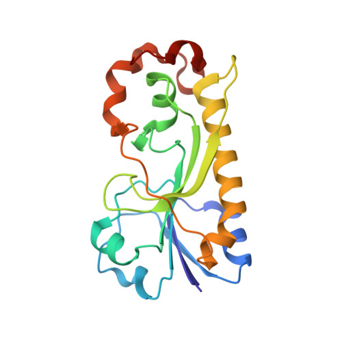
6CI2

Crystal structure of the formyltransferase PseJ from Anoxybacillus kamchatkensis


Experimental Data Snapshot

  • Method: X-RAY DIFFRACTION
  • Resolution: 1.96 Å
  • R-Value Free: 0.223 
  • R-Value Work: 0.180 
  • R-Value Observed: 0.184 

Starting Model: experimental
View more details

wwPDB Validation   3D Report Full Report


This is version 1.4 of the entry. See complete history


Literature

Structural Insight into a Novel Formyltransferase and Evolution to a Nonribosomal Peptide Synthetase Tailoring Domain.

Reimer, J.M.Harb, I.Ovchinnikova, O.G.Jiang, J.Whitfield, C.Schmeing, T.M.

(2018) ACS Chem Biol 13: 3161-3172

  • DOI: https://doi.org/10.1021/acschembio.8b00739
  • Primary Citation of Related Structures:  
    6CI2, 6CI4, 6CI5, 6EDK

  • PubMed Abstract: 

    Nonribosomal peptide synthetases (NRPSs) increase the chemical diversity of their products by acquiring tailoring domains. Linear gramicidin synthetase starts with a tailoring formylation (F) domain, which likely originated from a sugar formyltransferase (FT) gene. Here, we present studies on an Anoxybacillus kamchatkensis sugar FT representative of the prehorizontal gene transfer FT. Gene cluster analysis reveals that this FT acts on a UDP-sugar in a novel pathway for synthesis of a 7-formamido derivative of CMP-pseudaminic acid. We recapitulate the pathway up to and including the formylation step in vitro, experimentally demonstrating the role of the FT. We also present X-ray crystal structures of the FT alone and with ligands, which unveil contrasts with other structurally characterized sugar FTs and show close structural similarity with the F domain. The structures reveal insights into the adaptations that were needed to co-opt and evolve a sugar FT into a functional and useful NRPS domain.


  • Organizational Affiliation

    Department of Biochemistry , McGill University , Montréal , Québec H3G 0B1 , Canada.


Macromolecules
Find similar proteins by:  (by identity cutoff)  |  3D Structure
Entity ID: 1
MoleculeChains Sequence LengthOrganismDetailsImage
formyltransferase PseJ192Anoxybacillus ayderensis G10Mutation(s): 0 
Entity Groups  
Sequence Clusters30% Identity50% Identity70% Identity90% Identity95% Identity100% Identity
Sequence Annotations
Expand
  • Reference Sequence
Experimental Data & Validation

Experimental Data

  • Method: X-RAY DIFFRACTION
  • Resolution: 1.96 Å
  • R-Value Free: 0.223 
  • R-Value Work: 0.180 
  • R-Value Observed: 0.184 
  • Space Group: C 1 2 1
Unit Cell:
Length ( Å )Angle ( ˚ )
a = 95.63α = 90
b = 77.51β = 103.84
c = 41.73γ = 90
Software Package:
Software NamePurpose
PHENIXrefinement
MOSFLMdata reduction
Aimlessdata scaling
PHASERphasing

Structure Validation

View Full Validation Report


Entry History & Funding Information

Deposition Data


Funding OrganizationLocationGrant Number
Canadian Institutes of Health Research (CIHR)CanadaFDN-148472

Revision History  (Full details and data files)

  • Version 1.0: 2018-10-17
    Type: Initial release
  • Version 1.1: 2018-11-07
    Changes: Data collection, Database references
  • Version 1.2: 2018-11-28
    Changes: Data collection, Database references
  • Version 1.3: 2020-01-08
    Changes: Author supporting evidence
  • Version 1.4: 2023-10-04
    Changes: Data collection, Database references, Refinement description