6G7L | pdb_00006g7l

Retinal isomerization in bacteriorhodopsin revealed by a femtosecond X-ray laser: 8.3 ms state structure


Experimental Data Snapshot

  • Method: X-RAY DIFFRACTION
  • Resolution: 1.90 Å
  • R-Value Free: 
    0.272 (Depositor), 0.270 (DCC) 
  • R-Value Work: 
    0.224 (Depositor), 0.224 (DCC) 
  • R-Value Observed: 
    0.227 (Depositor) 

Starting Model: experimental
View more details

wwPDB Validation 3D Report Full Report

Validation slider image for 6G7L

Ligand Structure Quality Assessment 


This is version 1.4 of the entry. See complete history

Literature

Retinal isomerization in bacteriorhodopsin captured by a femtosecond x-ray laser.

Nogly, P.Weinert, T.James, D.Carbajo, S.Ozerov, D.Furrer, A.Gashi, D.Borin, V.Skopintsev, P.Jaeger, K.Nass, K.Bath, P.Bosman, R.Koglin, J.Seaberg, M.Lane, T.Kekilli, D.Brunle, S.Tanaka, T.Wu, W.Milne, C.White, T.Barty, A.Weierstall, U.Panneels, V.Nango, E.Iwata, S.Hunter, M.Schapiro, I.Schertler, G.Neutze, R.Standfuss, J.

(2018) Science 361

  • DOI: https://doi.org/10.1126/science.aat0094
  • Primary Citation Related Structures: 
    6G7H, 6G7I, 6G7J, 6G7K, 6G7L

  • PubMed Abstract: 

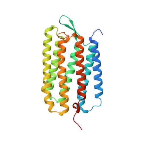
    Ultrafast isomerization of retinal is the primary step in photoresponsive biological functions including vision in humans and ion transport across bacterial membranes. We used an x-ray laser to study the subpicosecond structural dynamics of retinal isomerization in the light-driven proton pump bacteriorhodopsin. A series of structural snapshots with near-atomic spatial resolution and temporal resolution in the femtosecond regime show how the excited all-trans retinal samples conformational states within the protein binding pocket before passing through a twisted geometry and emerging in the 13-cis conformation. Our findings suggest ultrafast collective motions of aspartic acid residues and functional water molecules in the proximity of the retinal Schiff base as a key facet of this stereoselective and efficient photochemical reaction.


  • Organizational Affiliation
    • Division of Biology and Chemistry-Laboratory for Biomolecular Research, Paul Scherrer Institut, 5232 Villigen, Switzerland.

Macromolecule Content 

  • Total Structure Weight: 34.45 kDa 
  • Atom Count: 2,039 
  • Modeled Residue Count: 230 
  • Deposited Residue Count: 262 
  • Unique protein chains: 1

Macromolecules

Find similar proteins by:|  3D Structure
Entity ID: 1
MoleculeChains  Sequence LengthOrganismDetailsImage
Bacteriorhodopsin262Halobacterium salinarum NRC-1Mutation(s): 0 
Membrane Entity: Yes 
UniProt
Find proteins for P02945 (Halobacterium salinarum (strain ATCC 700922 / JCM 11081 / NRC-1))
Explore P02945 
Go to UniProtKB:  P02945
Entity Groups
Sequence Clusters30% Identity50% Identity70% Identity90% Identity95% Identity100% Identity
UniProt GroupP02945
Sequence Annotations
Expand
Reference Sequence

Small Molecules

Ligands 3 Unique
IDChains Name / Formula / InChI Key2D Diagram3D Interactions
LI1

Query on LI1


Download:Ideal Coordinates CCD File
C [auth A]
D [auth A]
E [auth A]
F [auth A]
G [auth A]
C [auth A],
D [auth A],
E [auth A],
F [auth A],
G [auth A],
H [auth A],
I [auth A]
1-[2,6,10.14-TETRAMETHYL-HEXADECAN-16-YL]-2-[2,10,14-TRIMETHYLHEXADECAN-16-YL]GLYCEROL
C42 H86 O3
YERVUJAKCNBGCR-BIHSMRAKSA-N
OLC

Query on OLC


Download:Ideal Coordinates CCD File
J [auth A],
K [auth A],
L [auth A],
M [auth A]
(2R)-2,3-dihydroxypropyl (9Z)-octadec-9-enoate
C21 H40 O4
RZRNAYUHWVFMIP-GDCKJWNLSA-N
RET

Query on RET


Download:Ideal Coordinates CCD File
B [auth A]RETINAL
C20 H28 O
NCYCYZXNIZJOKI-OVSJKPMPSA-N

Experimental Data & Validation

Experimental Data

  • Method: X-RAY DIFFRACTION
  • Resolution: 1.90 Å
  • R-Value Free:  0.272 (Depositor), 0.270 (DCC) 
  • R-Value Work:  0.224 (Depositor), 0.224 (DCC) 
  • R-Value Observed: 0.227 (Depositor) 
Space Group: P 63
Unit Cell:
Length ( Å )Angle ( ˚ )
a = 62.32α = 90
b = 62.32β = 90
c = 111.1γ = 120
Software Package:
Software NamePurpose
PHENIXrefinement
CrystFELdata reduction
CrystFELdata scaling
PHASERphasing

Structure Validation

View Full Validation Report


Ligand Structure Quality Assessment 


Entry History 

& Funding Information

Deposition Data


Funding OrganizationLocationGrant Number
Swiss National Science FoundationSwitzerland31003A_159558
Swiss National Science FoundationSwitzerland310030_153145
Swiss National Science FoundationSwitzerlandPZ00P3_174169
European CommissionSwitzerland290605

Revision History  (Full details and data files)

  • Version 1.0: 2018-06-27
    Type: Initial release
  • Version 1.1: 2018-07-25
    Changes: Data collection, Database references
  • Version 1.2: 2018-11-14
    Changes: Data collection
  • Version 1.3: 2024-01-17
    Changes: Data collection, Database references, Refinement description
  • Version 1.4: 2024-10-16
    Changes: Structure summary