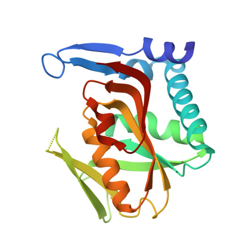
Structural basis for substrate selectivity and nucleophilic substitution mechanisms in human adenine phosphoribosyltransferase catalyzed reaction.
Ozeir, M., Huyet, J., Burgevin, M.C., Pinson, B., Chesney, F., Remy, J.M., Siddiqi, A.R., Lupoli, R., Pinon, G., Saint-Marc, C., Gibert, J.F., Morales, R., Ceballos-Picot, I., Barouki, R., Daignan-Fornier, B., Olivier-Bandini, A., Auge, F., Nioche, P.(2019) J Biological Chem 294: 11980-11991
- PubMed: 31160323
- DOI: https://doi.org/10.1074/jbc.RA119.009087
- Primary Citation of Related Structures:
6HGP, 6HGQ, 6HGR, 6HGS - PubMed Abstract:
The reversible adenine phosphoribosyltransferase enzyme (APRT) is essential for purine homeostasis in prokaryotes and eukaryotes. In humans, APRT (hAPRT) is the only enzyme known to produce AMP in cells from dietary adenine. APRT can also process adenine analogs, which are involved in plant development or neuronal homeostasis. However, the molecular mechanism underlying substrate specificity of APRT and catalysis in both directions of the reaction remains poorly understood. Here we present the crystal structures of hAPRT complexed to three cellular nucleotide analogs (hypoxanthine, IMP, and GMP) that we compare with the phosphate-bound enzyme. We established that binding to hAPRT is substrate shape-specific in the forward reaction, whereas it is base-specific in the reverse reaction. Furthermore, a quantum mechanics/molecular mechanics (QM/MM) analysis suggests that the forward reaction is mainly a nucleophilic substitution of type 2 (S N 2) with a mix of S N 1-type molecular mechanism. Based on our structural analysis, a magnesium-assisted S N 2-type mechanism would be involved in the reverse reaction. These results provide a framework for understanding the molecular mechanism and substrate discrimination in both directions by APRTs. This knowledge can play an instrumental role in the design of inhibitors, such as antiparasitic agents, or adenine-based substrates.
- Université Paris Descartes, Sorbonne Paris Cité, UFR des Sciences Fondamentales et Biomédicales, UMR 1124, Centre Interdisciplinaire Chimie Biologie-Paris, Paris, 75006, France; INSERM, UMR 1124, Paris, 75006, France.
Organizational Affiliation:


