6HN1 | pdb_00006hn1

Complex of Caprine Serum Albumin with diclofenac


Experimental Data Snapshot

  • Method: X-RAY DIFFRACTION
  • Resolution: 1.55 Å
  • R-Value Free: 
    0.239 (Depositor), 0.234 (DCC) 
  • R-Value Work: 
    0.165 (Depositor), 0.175 (DCC) 
  • R-Value Observed: 
    0.166 (Depositor) 

Starting Model: experimental
View more details

wwPDB Validation 3D Report Full Report

Validation slider image for 6HN1

Ligand Structure Quality Assessment 


This is version 1.3 of the entry. See complete history

Literature

Structural Investigation of Diclofenac Binding to Ovine, Caprine, and Leporine Serum Albumins.

Talaj, J.A.Zielinski, K.Bujacz, A.

(2023) Int J Mol Sci 24

  • DOI: https://doi.org/10.3390/ijms24021534
  • Primary Citation Related Structures: 
    6HN0, 6HN1, 8BSG

  • PubMed Abstract: 

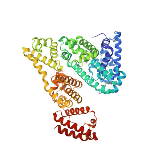
    Free drug concentration in the blood sera is crucial for its appropriate activity. Serum albumin, the universal blood carrier protein, is responsible for transporting drugs and releasing them into the bloodstream. Therefore, a drug's binding to SA is especially important for its bioavailability and it is a key problem in the drug design process. In this paper, we present crystal structures of three animal serum albumin complexes: ovine, caprine, and leporine, with diclofenac, a popular non-steroidal anti-inflammatory drug that is used in therapy of chronic and acute pain. Details of diclofenac binding mode by the presented serum albumins are compared with analogous complexes of human and equine serum albumins. The analysis of the occupied binding pockets in crystal structures of the investigated serum albumins from different mammals shows that they have two common and a number of unique diclofenac binding sites. The most intriguing is the fact that the albumins from the described species are able to bind different numbers of molecules of this popular anti-inflammatory drug, but none of the binding sites overlap with ones in the human serum albumin.


  • Organizational Affiliation
    • Department of Molecular Biology of Cancer, Medical University of Lodz, 6/8 Mazowiecka Street, 92-215 Lodz, Poland.

Macromolecule Content 

  • Total Structure Weight: 68.23 kDa 
  • Atom Count: 5,330 
  • Modeled Residue Count: 583 
  • Deposited Residue Count: 583 
  • Unique protein chains: 1

Macromolecules

Find similar proteins by:|  3D Structure
Entity ID: 1
MoleculeChains  Sequence LengthOrganismDetailsImage
Albumin583Capra hircusMutation(s): 0 
UniProt
Find proteins for A0A452F7K4 (Capra hircus)
Explore A0A452F7K4 
Go to UniProtKB:  A0A452F7K4
Entity Groups
Sequence Clusters30% Identity50% Identity70% Identity90% Identity95% Identity100% Identity
UniProt GroupA0A452F7K4
Sequence Annotations
Expand
Reference Sequence

Small Molecules

Ligands 2 Unique
IDChains Name / Formula / InChI Key2D Diagram3D Interactions
DIF
(Subject of Investigation/LOI)

Query on DIF


Download:Ideal Coordinates CCD File
B [auth A]
C [auth A]
D [auth A]
E [auth A]
F [auth A]
B [auth A],
C [auth A],
D [auth A],
E [auth A],
F [auth A],
G [auth A]
2-[2,6-DICHLOROPHENYL)AMINO]BENZENEACETIC ACID
C14 H11 Cl2 N O2
DCOPUUMXTXDBNB-UHFFFAOYSA-N
CL

Query on CL


Download:Ideal Coordinates CCD File
H [auth A]CHLORIDE ION
Cl
VEXZGXHMUGYJMC-UHFFFAOYSA-M

Experimental Data & Validation

Experimental Data

  • Method: X-RAY DIFFRACTION
  • Resolution: 1.55 Å
  • R-Value Free:  0.239 (Depositor), 0.234 (DCC) 
  • R-Value Work:  0.165 (Depositor), 0.175 (DCC) 
  • R-Value Observed: 0.166 (Depositor) 
Space Group: P 21 21 21
Unit Cell:
Length ( Å )Angle ( ˚ )
a = 41.96α = 90
b = 66.28β = 90
c = 212.58γ = 90
Software Package:
Software NamePurpose
REFMACrefinement
XDSdata reduction
XDSdata scaling
MOLREPphasing

Structure Validation

View Full Validation Report


Ligand Structure Quality Assessment 


Entry History 

& Funding Information

Deposition Data


Funding OrganizationLocationGrant Number
Poland2013/11/B/ST5/02271

Revision History  (Full details and data files)

  • Version 1.0: 2019-10-09
    Type: Initial release
  • Version 1.1: 2023-02-08
    Changes: Database references
  • Version 1.2: 2024-02-07
    Changes: Data collection, Refinement description
  • Version 1.3: 2024-11-20
    Changes: Structure summary