6I1I

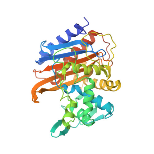
Crystal structure of TP domain from Escherichia coli penicillin-binding protein 3 in complex with penicillin


Experimental Data Snapshot

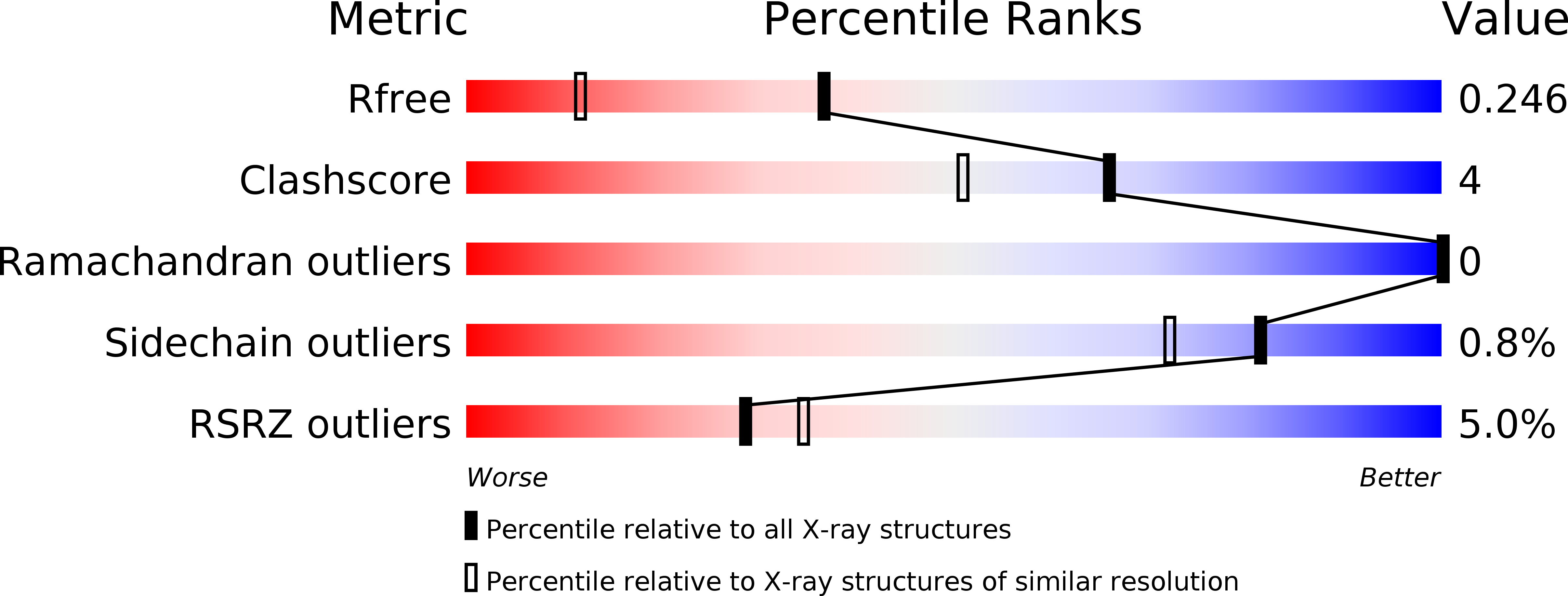
  • Method: X-RAY DIFFRACTION
  • Resolution: 1.75 Å
  • R-Value Free: 0.247 
  • R-Value Work: 0.220 
  • R-Value Observed: 0.222 

Starting Model: experimental
View more details

wwPDB Validation   3D Report Full Report


Ligand Structure Quality Assessment 


This is version 1.2 of the entry. See complete history


Literature

Novel and Improved Crystal Structures of H. influenzae, E. coli and P. aeruginosa Penicillin-Binding Protein 3 (PBP3) and N. gonorrhoeae PBP2: Toward a Better Understanding of beta-Lactam Target-Mediated Resistance.

Bellini, D.Koekemoer, L.Newman, H.Dowson, C.G.

(2019) J Mol Biol 431: 3501-3519

  • DOI: https://doi.org/10.1016/j.jmb.2019.07.010
  • Primary Citation of Related Structures:  
    6HZJ, 6HZO, 6HZQ, 6HZR, 6I1E, 6I1I, 6R3X, 6R40, 6R42

  • PubMed Abstract: 

    Even with the emergence of antibiotic resistance, penicillin and the wider family of β-lactams have remained the single most important family of antibiotics. The periplasmic/extra-cytoplasmic targets of penicillin are a family of enzymes with a highly conserved catalytic activity involved in the final stage of bacterial cell wall (peptidoglycan) biosynthesis. Named after their ability to bind penicillin, rather than their catalytic activity, these key targets are called penicillin-binding proteins (PBPs). Resistance is predominantly mediated by reducing the target drug concentration via β-lactamases; however, naturally transformable bacteria have also acquired target-mediated resistance by inter-species recombination. Here we focus on structural based interpretations of amino acid alterations associated with the emergence of resistance within clinical isolates and include new PBP3 structures along with new, and improved, PBP-β-lactam co-structures.


  • Organizational Affiliation

    School of Life Sciences, University of Warwick, Coventry, CV47AL, UK.


Macromolecules
Find similar proteins by:  (by identity cutoff)  |  3D Structure
Entity ID: 1
MoleculeChains Sequence LengthOrganismDetailsImage
Peptidoglycan D,D-transpeptidase FtsI,Peptidoglycan D,D-transpeptidase FtsI348Escherichia coli O157:H7Mutation(s): 0 
Gene Names: ftsIpbpBZ0094ECs0088
EC: 3.4.16.4
UniProt
Find proteins for P0AD68 (Escherichia coli (strain K12))
Explore P0AD68 
Go to UniProtKB:  P0AD68
Entity Groups  
Sequence Clusters30% Identity50% Identity70% Identity90% Identity95% Identity100% Identity
UniProt GroupP0AD68
Sequence Annotations
Expand
  • Reference Sequence
Small Molecules
Ligands 1 Unique
IDChains Name / Formula / InChI Key2D Diagram3D Interactions
JPP (Subject of Investigation/LOI)
Query on JPP

Download Ideal Coordinates CCD File 
B [auth A]Piperacillin (Open Form)
C23 H29 N5 O7 S
CJYMHKMECTVNSA-XLMAVXFVSA-N
Experimental Data & Validation

Experimental Data

  • Method: X-RAY DIFFRACTION
  • Resolution: 1.75 Å
  • R-Value Free: 0.247 
  • R-Value Work: 0.220 
  • R-Value Observed: 0.222 
  • Space Group: C 2 2 21
Unit Cell:
Length ( Å )Angle ( ˚ )
a = 99.714α = 90
b = 147.672β = 90
c = 43.254γ = 90
Software Package:
Software NamePurpose
REFMACrefinement
DIALSdata reduction
Aimlessdata scaling
MrBUMPphasing

Structure Validation

View Full Validation Report


Ligand Structure Quality Assessment 


Entry History & Funding Information

Deposition Data


Funding OrganizationLocationGrant Number
Medical Research Council (United Kingdom)United Kingdomgrant.MR/P007503/1

Revision History  (Full details and data files)

  • Version 1.0: 2019-11-20
    Type: Initial release
  • Version 1.1: 2020-02-19
    Changes: Database references
  • Version 1.2: 2024-01-24
    Changes: Data collection, Database references, Refinement description