6JFM | pdb_00006jfm

Mitofusin2 (MFN2)_T111D


Experimental Data Snapshot

  • Method: X-RAY DIFFRACTION
  • Resolution: 2.09 Å
  • R-Value Free: 
    0.229 (Depositor), 0.230 (DCC) 
  • R-Value Work: 
    0.176 (Depositor), 0.176 (DCC) 
  • R-Value Observed: 
    0.178 (Depositor) 

Starting Model: experimental
View more details

wwPDB Validation 3D Report Full Report

Validation slider image for 6JFM

This is version 1.2 of the entry. See complete history

Literature

Structural insights of human mitofusin-2 into mitochondrial fusion and CMT2A onset.

Li, Y.J.Cao, Y.L.Feng, J.X.Qi, Y.Meng, S.Yang, J.F.Zhong, Y.T.Kang, S.Chen, X.Lan, L.Luo, L.Yu, B.Chen, S.Chan, D.C.Hu, J.Gao, S.

(2019) Nat Commun 10: 4914-4914

  • DOI: https://doi.org/10.1038/s41467-019-12912-0
  • Primary Citation Related Structures: 
    6JFK, 6JFL, 6JFM

  • PubMed Abstract: 

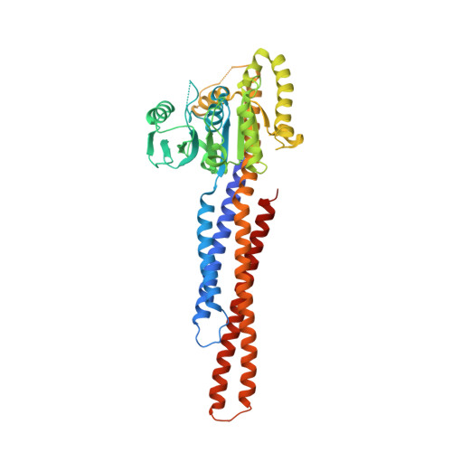
    Mitofusin-2 (MFN2) is a dynamin-like GTPase that plays a central role in regulating mitochondrial fusion and cell metabolism. Mutations in MFN2 cause the neurodegenerative disease Charcot-Marie-Tooth type 2A (CMT2A). The molecular basis underlying the physiological and pathological relevance of MFN2 is unclear. Here, we present crystal structures of truncated human MFN2 in different nucleotide-loading states. Unlike other dynamin superfamily members including MFN1, MFN2 forms sustained dimers even after GTP hydrolysis via the GTPase domain (G) interface, which accounts for its high membrane-tethering efficiency. The biochemical discrepancy between human MFN2 and MFN1 largely derives from a primate-only single amino acid variance. MFN2 and MFN1 can form heterodimers via the G interface in a nucleotide-dependent manner. CMT2A-related mutations, mapping to different functional zones of MFN2, lead to changes in GTP hydrolysis and homo/hetero-association ability. Our study provides fundamental insight into how mitofusins mediate mitochondrial fusion and the ways their disruptions cause disease.


  • Organizational Affiliation
    • State Key Laboratory of Oncology in South China, Collaborative Innovation Center for Cancer Medicine, Sun Yat-sen University Cancer Center, 510060, Guangzhou, China.

Macromolecule Content 

  • Total Structure Weight: 99.93 kDa 
  • Atom Count: 7,099 
  • Modeled Residue Count: 841 
  • Deposited Residue Count: 876 
  • Unique protein chains: 1

Macromolecules

Find similar proteins by:|  3D Structure
Entity ID: 1
MoleculeChains  Sequence LengthOrganismDetailsImage
Mitofusin-2,Mitofusin-2
A, B
438Homo sapiensMacaca mulatta
This entity is chimeric
Mutation(s): 1 
Gene Names: MFN2CPRP1KIAA0214EGK_00242
EC: 3.6.5
UniProt & NIH Common Fund Data Resources
Find proteins for O95140 (Homo sapiens)
Explore O95140 
Go to UniProtKB:  O95140
PHAROS:  O95140
GTEx:  ENSG00000116688 
Entity Groups
Sequence Clusters30% Identity50% Identity70% Identity90% Identity95% Identity100% Identity
UniProt GroupO95140
Sequence Annotations
Expand
Reference Sequence

Experimental Data & Validation

Experimental Data

  • Method: X-RAY DIFFRACTION
  • Resolution: 2.09 Å
  • R-Value Free:  0.229 (Depositor), 0.230 (DCC) 
  • R-Value Work:  0.176 (Depositor), 0.176 (DCC) 
  • R-Value Observed: 0.178 (Depositor) 
Space Group: P 1 21 1
Unit Cell:
Length ( Å )Angle ( ˚ )
a = 44.437α = 90
b = 126.563β = 102.46
c = 79.972γ = 90
Software Package:
Software NamePurpose
PHENIXrefinement
REFMACrefinement
XDSdata reduction
XSCALEdata scaling
MOLREPphasing

Structure Validation

View Full Validation Report


Entry History 

& Funding Information

Deposition Data


Funding OrganizationLocationGrant Number
National Natural Science Foundation of ChinaChina--

Revision History  (Full details and data files)

  • Version 1.0: 2019-11-13
    Type: Initial release
  • Version 1.1: 2023-11-22
    Changes: Data collection, Database references, Derived calculations, Refinement description
  • Version 1.2: 2024-10-30
    Changes: Structure summary