6KG1

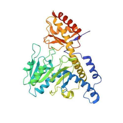
NifS from Helicobacter pylori, soaked with L-cysteine for 180 sec


Experimental Data Snapshot

  • Method: X-RAY DIFFRACTION
  • Resolution: 2.70 Å
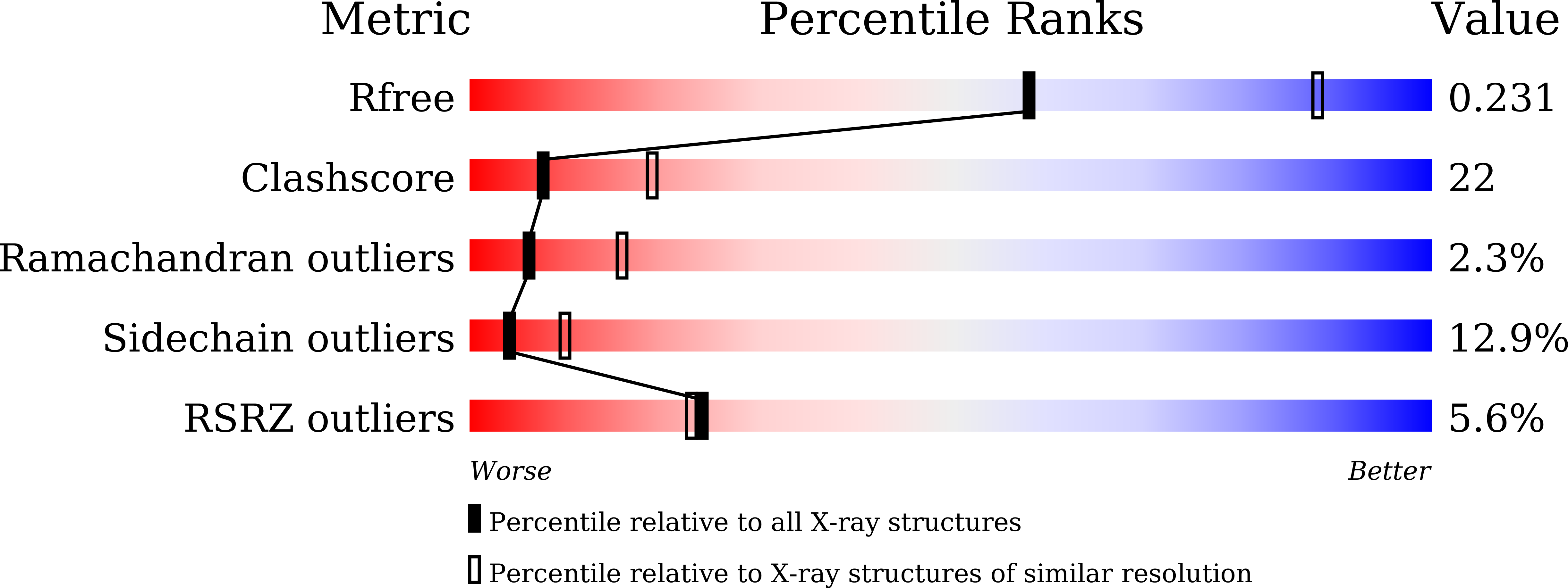
  • R-Value Free: 0.231 
  • R-Value Work: 0.180 
  • R-Value Observed: 0.183 

Starting Model: experimental
View more details

wwPDB Validation   3D Report Full Report


Ligand Structure Quality Assessment 


This is version 1.3 of the entry. See complete history


Literature

Snapshots of PLP-substrate and PLP-product external aldimines as intermediates in two types of cysteine desulfurase enzymes.

Nakamura, R.Hikita, M.Ogawa, S.Takahashi, Y.Fujishiro, T.

(2020) FEBS J 287: 1138-1154

  • DOI: https://doi.org/10.1111/febs.15081
  • Primary Citation of Related Structures:  
    5WT2, 5WT4, 5ZS9, 5ZSK, 5ZSO, 5ZSP, 5ZST, 6KFZ, 6KG0, 6KG1

  • PubMed Abstract: 

    Cysteine desulfurase enzymes catalyze sulfur mobilization from l-cysteine to sulfur-containing biomolecules such as iron-sulfur (Fe-S) clusters and thio-tRNAs. The enzymes utilize the cofactor pyridoxal-5'-phosphate (PLP), which forms the external substrate- and product-aldimines and ketimines during catalysis and are grouped into two types (I and II) based on their different catalytic loops. To clarify the structure-based catalytic mechanisms for each group, we determined the structures of the external substrate- and product-aldimines as catalytic intermediates of NifS (type I) and SufS (type II) that are involved in Fe-S cluster biosynthesis using X-ray crystallographic snapshot analysis. As a common intermediate structure, the thiol group of the PLP-l-cysteine external aldimine is stabilized by the conserved histidine adjacent to PLP through a polar interaction. This interaction makes the thiol group orientated for subsequent nucleophilic attack by a conserved cysteine residue on the catalytic loop in the state of PLP-l-cysteine ketimine, which is formed from the PLP-l-cysteine aldimine. Unlike the intermediates, structural changes of the loops were different between the type I and II enzymes. In the type I enzyme, conformational and topological change of the loop is necessary for nucleophilic attack by the cysteine. In contrast, the loop in type II cysteine desulfurase enzymes showed no large conformational change; rather, it might possibly orient the thiol group of the catalytic cysteine for nucleophilic attack toward PLP-l-cysteine. The present structures allow a revision of the catalytic mechanism and may provide a clue for consideration of enzyme function, structural diversity, and evolution of cysteine desulfurase enzymes. DATABASE: Structural data are available in PDB database under the accession numbers 5WT2, 5WT4, 5ZSP, 5ZST, 5ZS9, 5ZSK, 5ZSO, 6KFZ, 6KG0, and 6KG1.


  • Organizational Affiliation

    Department of Biochemistry and Molecular Biology, Graduate School of Science and Engineering, Saitama University, Saitama, Japan.


Macromolecules
Find similar proteins by:  (by identity cutoff)  |  3D Structure
Entity ID: 1
MoleculeChains Sequence LengthOrganismDetailsImage
Cysteine desulfurase IscS401Helicobacter pylori 26695Mutation(s): 2 
Gene Names: iscSHP_0220
EC: 2.8.1.7
UniProt
Find proteins for O25008 (Helicobacter pylori (strain ATCC 700392 / 26695))
Explore O25008 
Go to UniProtKB:  O25008
Entity Groups  
Sequence Clusters30% Identity50% Identity70% Identity90% Identity95% Identity100% Identity
UniProt GroupO25008
Sequence Annotations
Expand
  • Reference Sequence
Small Molecules
Ligands 3 Unique
IDChains Name / Formula / InChI Key2D Diagram3D Interactions
PDA
Query on PDA

Download Ideal Coordinates CCD File 
D [auth A]2-[(3-HYDROXY-2-METHYL-5-PHOSPHONOOXYMETHYL-PYRIDIN-4-YLMETHYL)-AMINO]-PROPIONIC ACID
C11 H17 N2 O7 P
WACJCHFWJNNBPR-ZETCQYMHSA-N
IPA
Query on IPA

Download Ideal Coordinates CCD File 
B [auth A]ISOPROPYL ALCOHOL
C3 H8 O
KFZMGEQAYNKOFK-UHFFFAOYSA-N
CL
Query on CL

Download Ideal Coordinates CCD File 
C [auth A]CHLORIDE ION
Cl
VEXZGXHMUGYJMC-UHFFFAOYSA-M
Experimental Data & Validation

Experimental Data

  • Method: X-RAY DIFFRACTION
  • Resolution: 2.70 Å
  • R-Value Free: 0.231 
  • R-Value Work: 0.180 
  • R-Value Observed: 0.183 
  • Space Group: P 41 21 2
Unit Cell:
Length ( Å )Angle ( ˚ )
a = 102.9α = 90
b = 102.9β = 90
c = 133.2γ = 90
Software Package:
Software NamePurpose
XSCALEdata scaling
REFMACrefinement
PDB_EXTRACTdata extraction
XDSdata reduction
MOLREPphasing

Structure Validation

View Full Validation Report


Ligand Structure Quality Assessment 


Entry History & Funding Information

Deposition Data


Funding OrganizationLocationGrant Number
Japan Society for the Promotion of ScienceJapan17K14510

Revision History  (Full details and data files)

  • Version 1.0: 2019-10-16
    Type: Initial release
  • Version 1.1: 2019-10-23
    Changes: Data collection, Database references
  • Version 1.2: 2020-04-01
    Changes: Database references
  • Version 1.3: 2023-11-22
    Changes: Data collection, Database references, Refinement description