6LFD | pdb_00006lfd

Crystal structure of VMB-1 at pH5.5(Bis-Tris)


Experimental Data Snapshot

  • Method: X-RAY DIFFRACTION
  • Resolution: 1.92 Å
  • R-Value Free: 
    0.239 (Depositor), 0.239 (DCC) 
  • R-Value Work: 
    0.200 (Depositor), 0.209 (DCC) 
  • R-Value Observed: 
    0.202 (Depositor) 

Starting Model: experimental
View more details

wwPDB Validation 3D Report Full Report

Validation slider image for 6LFD

Ligand Structure Quality Assessment 


This is version 1.2 of the entry. See complete history

Literature

Crystal structure of VMB-1 at pH 5.5(Bis-Tris)

Chen, S.Cheng, Q.

To be published.

Macromolecule Content 

  • Total Structure Weight: 103.53 kDa 
  • Atom Count: 7,203 
  • Modeled Residue Count: 877 
  • Deposited Residue Count: 936 
  • Unique protein chains: 1

Macromolecules

Find similar proteins by:|  3D Structure
Entity ID: 1
MoleculeChains  Sequence LengthOrganismDetailsImage
VMB-1
A, B, C, D
234Vibrio alginolyticusMutation(s): 0 
Gene Names: vmb-1
EC: 3.5.2.6
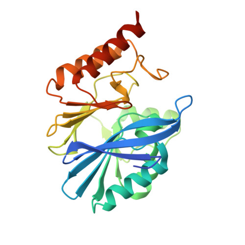
UniProt
Find proteins for A0A5Q5ADH9 (Vibrio alginolyticus)
Explore A0A5Q5ADH9 
Go to UniProtKB:  A0A5Q5ADH9
Entity Groups
Sequence Clusters30% Identity50% Identity70% Identity90% Identity95% Identity100% Identity
UniProt GroupA0A5Q5ADH9
Sequence Annotations
Expand
Reference Sequence

Small Molecules

Ligands 2 Unique
IDChains Name / Formula / InChI Key2D Diagram3D Interactions
GOL
(Subject of Investigation/LOI)

Query on GOL


Download:Ideal Coordinates CCD File
I [auth B],
J [auth B]
GLYCEROL
C3 H8 O3
PEDCQBHIVMGVHV-UHFFFAOYSA-N
ZN
(Subject of Investigation/LOI)

Query on ZN


Download:Ideal Coordinates CCD File
E [auth A]
F [auth A]
G [auth B]
H [auth B]
K [auth C]
E [auth A],
F [auth A],
G [auth B],
H [auth B],
K [auth C],
L [auth C],
M [auth D],
N [auth D]
ZINC ION
Zn
PTFCDOFLOPIGGS-UHFFFAOYSA-N

Experimental Data & Validation

Experimental Data

  • Method: X-RAY DIFFRACTION
  • Resolution: 1.92 Å
  • R-Value Free:  0.239 (Depositor), 0.239 (DCC) 
  • R-Value Work:  0.200 (Depositor), 0.209 (DCC) 
  • R-Value Observed: 0.202 (Depositor) 
Space Group: P 21 21 21
Unit Cell:
Length ( Å )Angle ( ˚ )
a = 73.11α = 90
b = 97.11β = 90
c = 132.66γ = 90
Software Package:
Software NamePurpose
REFMACrefinement
xia2data scaling
PDB_EXTRACTdata extraction
xia2data reduction
PHASERphasing

Structure Validation

View Full Validation Report


Ligand Structure Quality Assessment 


Entry History 

Deposition Data

Revision History  (Full details and data files)

  • Version 1.0: 2020-02-26
    Type: Initial release
  • Version 1.1: 2020-03-18
    Changes: Database references, Source and taxonomy, Structure summary
  • Version 1.2: 2023-11-22
    Changes: Data collection, Database references, Derived calculations, Refinement description