6LFE | pdb_00006lfe

Rat-COMT, Nitecapone,SAM and Mg bond


Experimental Data Snapshot

  • Method: X-RAY DIFFRACTION
  • Resolution: 1.60 Å
  • R-Value Free: 
    0.166 (Depositor), 0.169 (DCC) 
  • R-Value Work: 
    0.147 (Depositor), 0.149 (DCC) 
  • R-Value Observed: 
    0.148 (Depositor) 

Starting Model: experimental
View more details

wwPDB Validation 3D Report Full Report

Validation slider image for 6LFE

Ligand Structure Quality Assessment 


This is version 1.3 of the entry. See complete history

Literature

Crystal Structure of Catechol O-Methyltransferase Complexed with Nitecapone.

Iijima, H.Takebe, K.Suzuki, M.Kobayashi, H.Takamiya, T.Saito, H.Niwa, N.Kuwada-Kusunose, T.

(2020) Chem Pharm Bull (Tokyo) 68: 447-451

  • DOI: https://doi.org/10.1248/cpb.c20-00011
  • Primary Citation Related Structures: 
    6LFE

  • PubMed Abstract: 

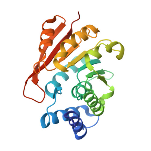
    Catechol O-methyltransferase (COMT) is known as an important drug-target protein in the field of Parkinson's disease. All clinically approved COMT inhibitors bring a 5-substituted-3-nitrocatechol ring as a pharmacophore, and they bind to COMT with S-adenosylmethionine (SAM) and an Mg 2+ ion to form a quaternary complex (COMT/SAM/Mg 2+ /inhibitor). However, structural information about such quaternary complexes is only available for a few inhibitors. Here, a new crystal structure of COMT complexed with nitecapone (5), SAM and Mg 2+ is revealed. Comparison of the structures of these complexes indicates that conformation of the catechol binding pocket is almost constant regardless of structure of the inhibitors. The only restriction of the side chain of inhibitors (i.e., the substituent at the 5-position of 3-nitrocatechol) seems to be that it does not make steric repulsion with COMT. However, recent crystallographic and biochemical studies suggest that COMT is a flexible protein, and its conformational flexibility seems crucial for its catalytic process. Based on this information, implications of these quaternary inhibitor complexes were investigated. Met 40 in the α2α3-loop makes atomic contacts with SAM or S-adenosylhomocysteine and the 3-position of the catechol inhibitor. This interaction seems to play a critical role in the affinity of the inhibitor and to stabilize the COMT/SAM/Mg 2+ /nitrocatechol inhibitor complex by fixing the flexible α2α3-loop.


  • Organizational Affiliation
    • School of Pharmacy, Nihon University.

Macromolecule Content 

  • Total Structure Weight: 25.94 kDa 
  • Atom Count: 2,006 
  • Modeled Residue Count: 213 
  • Deposited Residue Count: 223 
  • Unique protein chains: 1

Macromolecules

Find similar proteins by:|  3D Structure
Entity ID: 1
MoleculeChains  Sequence LengthOrganismDetailsImage
Catechol O-methyltransferase223Rattus norvegicusMutation(s): 0 
Gene Names: Comt
EC: 2.1.1.6
UniProt
Find proteins for P22734 (Rattus norvegicus)
Explore P22734 
Go to UniProtKB:  P22734
Entity Groups
Sequence Clusters30% Identity50% Identity70% Identity90% Identity95% Identity100% Identity
UniProt GroupP22734
Sequence Annotations
Expand
Reference Sequence

Small Molecules

Ligands 5 Unique
IDChains Name / Formula / InChI Key2D Diagram3D Interactions
SAM
(Subject of Investigation/LOI)

Query on SAM


Download:Ideal Coordinates CCD File
B [auth A]S-ADENOSYLMETHIONINE
C15 H22 N6 O5 S
MEFKEPWMEQBLKI-FCKMPRQPSA-N
EAO
(Subject of Investigation/LOI)

Query on EAO


Download:Ideal Coordinates CCD File
C [auth A]3-(3,4-dihydroxy-5-nitrobenzylidene)pentane-2,4-dione
C12 H11 N O6
UPMRZALMHVUCIN-UHFFFAOYSA-N
PEG

Query on PEG


Download:Ideal Coordinates CCD File
G [auth A],
H [auth A]
DI(HYDROXYETHYL)ETHER
C4 H10 O3
MTHSVFCYNBDYFN-UHFFFAOYSA-N
IPA

Query on IPA


Download:Ideal Coordinates CCD File
E [auth A],
F [auth A]
ISOPROPYL ALCOHOL
C3 H8 O
KFZMGEQAYNKOFK-UHFFFAOYSA-N
MG
(Subject of Investigation/LOI)

Query on MG


Download:Ideal Coordinates CCD File
D [auth A]MAGNESIUM ION
Mg
JLVVSXFLKOJNIY-UHFFFAOYSA-N

Experimental Data & Validation

Experimental Data

  • Method: X-RAY DIFFRACTION
  • Resolution: 1.60 Å
  • R-Value Free:  0.166 (Depositor), 0.169 (DCC) 
  • R-Value Work:  0.147 (Depositor), 0.149 (DCC) 
  • R-Value Observed: 0.148 (Depositor) 
Space Group: P 21 21 21
Unit Cell:
Length ( Å )Angle ( ˚ )
a = 49.665α = 90
b = 53.298β = 90
c = 80.786γ = 90
Software Package:
Software NamePurpose
PHENIXrefinement
XDSdata reduction
Aimlessdata scaling
MOLREPphasing
PDB_EXTRACTdata extraction

Structure Validation

View Full Validation Report


Ligand Structure Quality Assessment 


Entry History 

& Funding Information

Deposition Data


Funding OrganizationLocationGrant Number
Japan Society for the Promotion of Science (JSPS)JapanJP15K08034
Japan Agency for Medical Research and Development (AMED)JapanJP18am0101001 (0318)

Revision History  (Full details and data files)

  • Version 1.0: 2020-03-04
    Type: Initial release
  • Version 1.1: 2022-03-02
    Changes: Database references
  • Version 1.2: 2022-03-09
    Changes: Derived calculations, Structure summary
  • Version 1.3: 2023-11-22
    Changes: Data collection, Refinement description