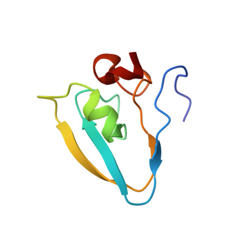
Use of the LC3B-fusion technique for biochemical and structural studies of proteins involved in the N-degron pathway.
Kim, L., Kwon, D.H., Heo, J., Park, M.R., Song, H.K.(2020) J Biological Chem 295: 2590-2600
- PubMed: 31919097
- DOI: https://doi.org/10.1074/jbc.RA119.010912
- Primary Citation of Related Structures:
6KGI, 6KGJ, 6KHZ, 6LHN - PubMed Abstract:
The N-degron pathway, formerly the N-end rule pathway, is a protein degradation process that determines the half-life of proteins based on their N-terminal residues. In contrast to the well-established in vivo studies over decades, in vitro studies of this pathway, including biochemical characterization and high-resolution structures, are relatively limited. In this study, we have developed a unique fusion technique using microtubule-associated protein 1A/1B light chain 3B, a key marker protein of autophagy, to tag the N terminus of the proteins involved in the N-degron pathway, which enables high yield of homogeneous target proteins with variable N-terminal residues for diverse biochemical studies including enzymatic and binding assays and substrate identification. Intriguingly, crystallization showed a markedly enhanced probability, even for the N-degron complexes. To validate our results, we determined the structures of select proteins in the N-degron pathway and compared them with the Protein Data Bank-deposited proteins. Furthermore, several biochemical applications of this technique were introduced. Therefore, this technique can be used as a general tool for the in vitro study of the N-degron pathway.
- Department of Life Sciences, Korea University, 145 Anam-ro, Seongbuk-gu, Seoul 02841, South Korea.
Organizational Affiliation:


