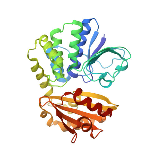
6LL8 | pdb_00006ll8

Type II inorganic pyrophosphatase (PPase) from the psychrophilic bacterium Shewanella sp. AS-11, Mg-PNP form


Experimental Data Snapshot

  • Method: X-RAY DIFFRACTION
  • Resolution: 1.30 Å
  • R-Value Free: 
    0.122 (Depositor), 0.120 (DCC) 
  • R-Value Work: 
    0.097 (Depositor), 0.100 (DCC) 

Starting Model: experimental
View more details

wwPDB Validation   3D Report Full Report


Ligand Structure Quality Assessment 

Created with Raphaël 2.3.0Worse 01 BetterLigand structure goodness of fit to experimental dataBest fitted 2PNClick on this verticalbar to view details

This is version 1.1 of the entry. See complete history


Literature

X-ray Crystallography and Electron Paramagnetic Resonance Spectroscopy Reveal Active Site Rearrangement of Cold-Adapted Inorganic Pyrophosphatase.

Horitani, M.Kusubayashi, K.Oshima, K.Yato, A.Sugimoto, H.Watanabe, K.

(2020) Sci Rep 10: 4368-4368

  • DOI: https://doi.org/10.1038/s41598-020-61217-6
  • Primary Citation of Related Structures:  
    6LL7, 6LL8

  • PubMed Abstract: 

    Inorganic pyrophosphatase (PPase) catalyses the hydrolysis reaction of inorganic pyrophosphate to phosphates. Our previous studies showed that manganese (Mn) activated PPase from the psychrophilic bacterium Shewanella sp. AS-11 (Mn-Sh-PPase) has a characteristic temperature dependence of the activity with an optimum at 5 °C. Here we report the X-ray crystallography and electron paramagnetic resonance (EPR) spectroscopy structural analyses of Sh-PPase in the absence and presence of substrate analogues. We successfully determined the crystal structure of Mn-Sh-PPase without substrate and Mg-activated Sh-PPase (Mg-Sh-PPase) complexed with substrate analogue (imidodiphosphate; PNP). Crystallographic studies revealed a bridged water placed at a distance from the di-Mn centre in Mn-Sh-PPase without substrate. The water came closer to the metal centre when PNP bound. EPR analysis of Mn-Sh-PPase without substrate revealed considerably weak exchange coupling, whose magnitude was increased by binding of substrate analogues. The data indicate that the bridged molecule has weak bonds with the di-Mn centre, which suggests a 'loose' structure, whereas it comes closer to di-Mn centre by substrate binding, which suggests a 'well-tuned' structure for catalysis. Thus, we propose that Sh-PPase can rearrange the active site and that the 'loose' structure plays an important role in the cold adaptation mechanism.


  • Organizational Affiliation

    Department of Applied Biochemistry and Food Science, Saga University, 1 Honjo-machi, Saga, Saga, 840-8502, Japan. horitani@cc.saga-u.ac.jp.


Macromolecules
Find similar proteins by:  (by identity cutoff)  |  3D Structure
Entity ID: 1
MoleculeChains Sequence LengthOrganismDetailsImage
Inorganic pyrophosphatase
A, B
309Shewanella sp. AS-11Mutation(s): 0 
Gene Names: ppia
EC: 3.6.1.1
UniProt
Find proteins for L8AXY8 (Shewanella sp. AS-11)
Explore L8AXY8 
Go to UniProtKB:  L8AXY8
Entity Groups  
Sequence Clusters30% Identity50% Identity70% Identity90% Identity95% Identity100% Identity
UniProt GroupL8AXY8
Sequence Annotations
Expand
  • Reference Sequence
Small Molecules
Ligands 6 Unique
IDChains Name / Formula / InChI Key2D Diagram3D Interactions
EPE
Query on EPE

Download Ideal Coordinates CCD File 
J [auth A],
T [auth B]
4-(2-HYDROXYETHYL)-1-PIPERAZINE ETHANESULFONIC ACID
C8 H18 N2 O4 S
JKMHFZQWWAIEOD-UHFFFAOYSA-N
2PN (Subject of Investigation/LOI)
Query on 2PN

Download Ideal Coordinates CCD File 
I [auth A],
S [auth B]
IMIDODIPHOSPHORIC ACID
H5 N O6 P2
GNGSOPFGGKKDQP-UHFFFAOYSA-N
GOL
Query on GOL

Download Ideal Coordinates CCD File 
K [auth A],
L [auth A],
U [auth B],
V [auth B]
GLYCEROL
C3 H8 O3
PEDCQBHIVMGVHV-UHFFFAOYSA-N
CA
Query on CA

Download Ideal Coordinates CCD File 
H [auth A]CALCIUM ION
Ca
BHPQYMZQTOCNFJ-UHFFFAOYSA-N
MG
Query on MG

Download Ideal Coordinates CCD File 
C [auth A]
D [auth A]
E [auth A]
F [auth A]
G [auth A]
MAGNESIUM ION
Mg
JLVVSXFLKOJNIY-UHFFFAOYSA-N
F
Query on F

Download Ideal Coordinates CCD File 
M [auth A],
W [auth B]
FLUORIDE ION
F
KRHYYFGTRYWZRS-UHFFFAOYSA-M
Experimental Data & Validation

Experimental Data

  • Method: X-RAY DIFFRACTION
  • Resolution: 1.30 Å
  • R-Value Free:  0.122 (Depositor), 0.120 (DCC) 
  • R-Value Work:  0.097 (Depositor), 0.100 (DCC) 
Space Group: P 1 21 1
Unit Cell:
Length ( Å )Angle ( ˚ )
a = 53.49α = 90
b = 78.98β = 98.593
c = 75.3γ = 90
Software Package:
Software NamePurpose
XDSdata scaling
REFMACrefinement
PDB_EXTRACTdata extraction
PHASERphasing
Cootmodel building
XDSdata reduction

Structure Validation

View Full Validation Report


Ligand Structure Quality Assessment 

Created with Raphaël 2.3.0Worse 01 BetterLigand structure goodness of fit to experimental dataBest fitted 2PNClick on this verticalbar to view details

Entry History & Funding Information

Deposition Data


Funding OrganizationLocationGrant Number
Japan Society for the Promotion of Science (JSPS)Japan17H06955
Japan Society for the Promotion of Science (JSPS)Japan18H02412

Revision History  (Full details and data files)

  • Version 1.0: 2020-03-25
    Type: Initial release
  • Version 1.1: 2023-11-22
    Changes: Data collection, Database references, Derived calculations, Refinement description